Lyu Jian, Wang Ju-Bo, Quan Yu, Gong Shouping
Neurosurgical Department, Second Affiliated Hospital, Xi'an Jiaotong University, Xi'an, 710004, China.
J Med Case Rep. 2019 Jul 21;13(1):222. doi: 10.1186/s13256-019-2158-9.
Management of the disproportionately large communicating fourth ventricle is still problematic.
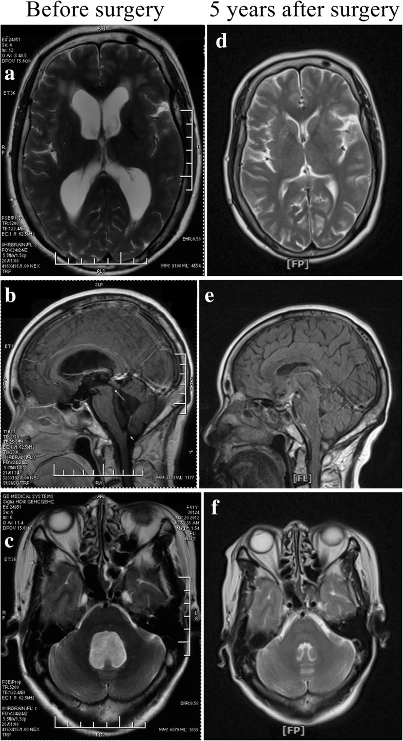
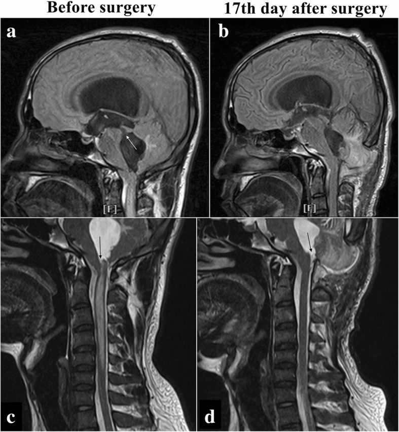
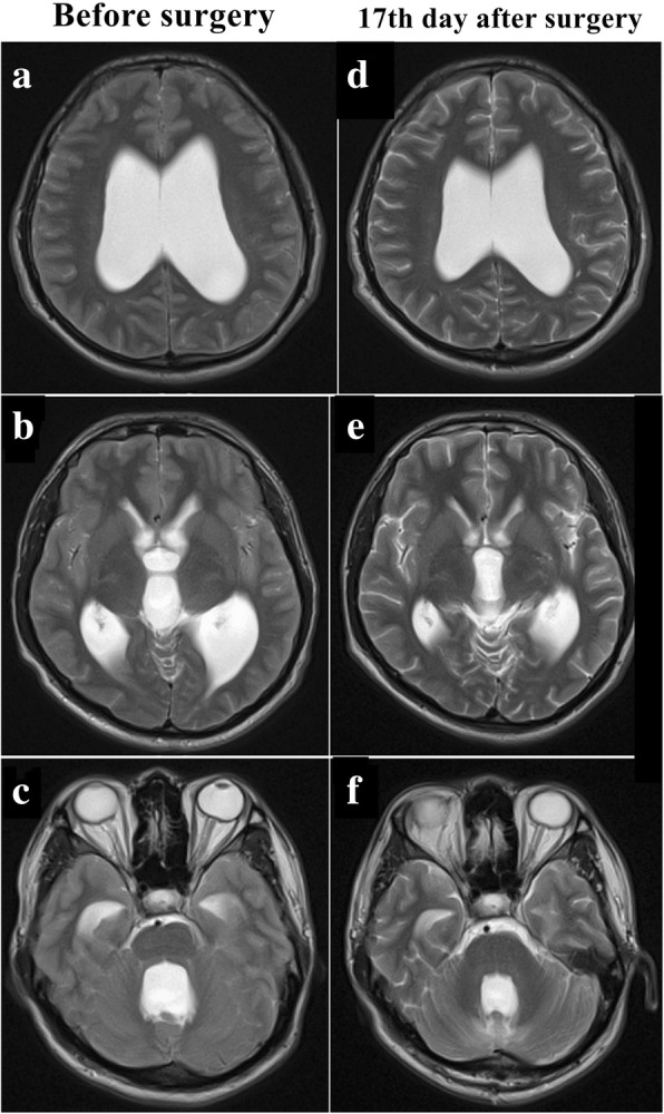
Two cases of disproportionately large communicating fourth ventricle were treated successfully. One was a case of a 51-year-old Han Chinese woman with a complaint of headache and dizziness of 1 year's duration. Magnetic resonance imaging (MRI) demonstrated hydrocephalus with a disproportionately large fourth ventricle. She underwent a ventriculo-peritoneal shunt of the right lateral ventricle. Her symptoms were relieved totally. Five years later, magnetic resonance imaging showed she had a normal ventricular system. The other case was a 24-year-old Han Chinese man with a 2-month history of headache and dizziness accompanied by progressive loss of bilateral vision. Magnetic resonance imaging revealed hydrocephalus with a disproportionately large fourth ventricle, crowded posterior cranial fossa, and syringomyelia extending from C1 to C5. He underwent suboccipital and C1 decompression and duraplasty. Shortly after the surgery, his symptoms were relieved completely, the syringomyelia completely disappeared, and the fourth ventricle became significantly smaller.
The management of the disproportionately large communicating fourth ventricle should be individualized. If it coexists with crowded posterior cranial fossa or syringomyelia, posterior fossa decompression could be an option for initial management. If there is no sign of crowded posterior cranial fossa or syringomyelia, shunt of the lateral ventricles might be the first choice.
不成比例增大的交通性第四脑室的管理仍然存在问题。
成功治疗了两例不成比例增大的交通性第四脑室病例。一例是一名51岁的汉族女性,主诉头痛和头晕1年。磁共振成像(MRI)显示脑积水伴不成比例增大的第四脑室。她接受了右侧脑室-腹腔分流术。她的症状完全缓解。五年后,磁共振成像显示她的脑室系统正常。另一例是一名24岁的汉族男性,有2个月的头痛和头晕病史,伴有双侧视力进行性丧失。磁共振成像显示脑积水伴不成比例增大的第四脑室、后颅窝拥挤以及从C1至C5的脊髓空洞症。他接受了枕下和C1减压及硬脑膜成形术。手术后不久,他的症状完全缓解,脊髓空洞症完全消失,第四脑室明显变小。
不成比例增大的交通性第四脑室的管理应个体化。如果它与后颅窝拥挤或脊髓空洞症并存,后颅窝减压可能是初始管理的一种选择。如果没有后颅窝拥挤或脊髓空洞症的迹象,侧脑室分流可能是首选。