Department of Critical Care Medicine, Shengjing Hospital of China Medical University, Shenyang, China.
Front Immunol. 2019 Jul 2;10:1491. doi: 10.3389/fimmu.2019.01491. eCollection 2019.
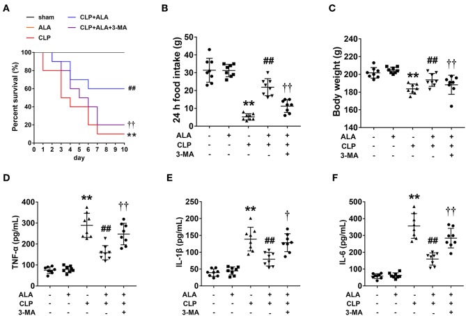
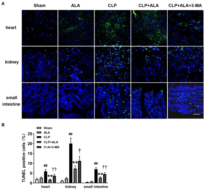
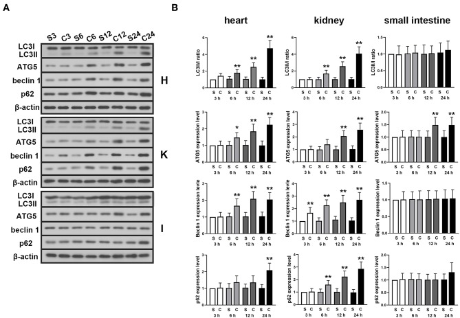
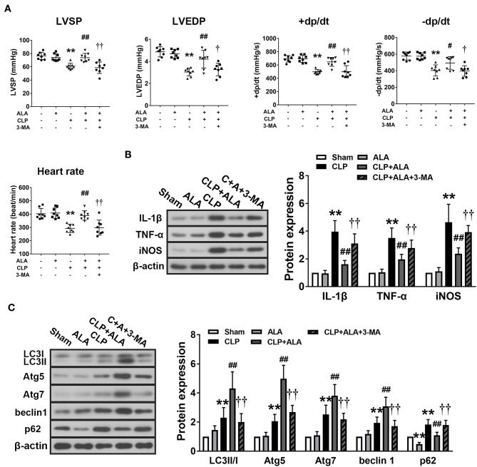
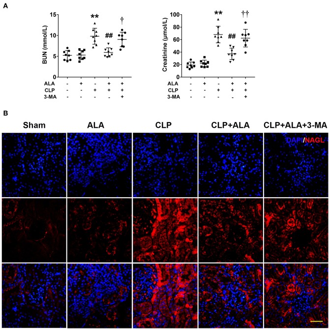
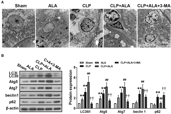
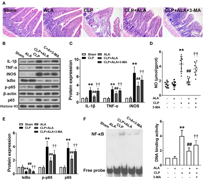
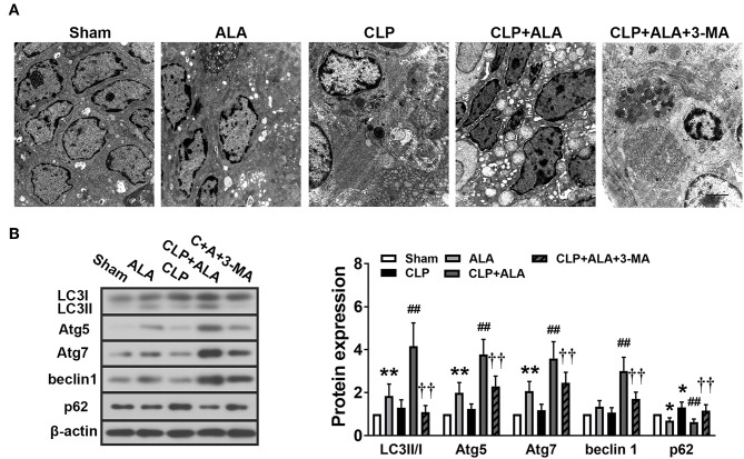
Alpha-lipoic acid (ALA) reportedly has protective effects against sepsis, which is a leading cause of mortality worldwide and is associated with multiple organ dysfunction. The present study aimed to investigate further the possible action mechanisms of ALA. Male Sprague-Dawley rats were subjected to cecal ligation and puncture (CLP) in order to establish a sepsis model. The rats received an oral gavage of 200 mg/kg ALA or saline immediately after surgery. The heart rate (HR), left ventricular systolic pressure (LVSP), left ventricular end-diastolic pressure (LVEDP) and maximum rising and lowering rates of left ventricular pressure (±dp/dt) were examined for assessing the cardiac function. Blood urea nitrogen (BUN) and serum creatinine levels were assessed for evaluating renal function. Neutrophil gelatinase-associated lipocalin (NAGL) was examined for reflecting acute renal injury. Histopathological alterations of the small intestine were examined by hematoxylin-eosin staining. The ultrastructure of the small intestine and kidney was observed under electron microscopy. The levels of autophagy- and inflammation-associated proteins were determined via western blot analysis. The binding of nuclear factor-kappa B (NF-κB) to DNA was tested via an electrophoretic mobility shift assay. Cell apoptosis was examined using TUNEL staining. ALA treatment improved the survival rate, restored the loss of body weight and pro-inflammatory cytokines production in the serum of CLP-induced septic rats. ALA improved the cardiac and renal functions, downregulated the expression levels of interleukin-1β, tumor necrosis factor-α, and inducible nitric oxide synthase in the myocardium and small intestine of septic rats. ALA treatment also inactivated the NF-κB signaling pathway in the small intestine. An examination of autophagy showed that ALA increased the LC3II/I ratio, upregulated Atg5, Atg7, and beclin-1 and downregulated p62 protein levels in the myocardium, kidney, and small intestine of septic rats, and further promoted autophagosome accumulation in the kidney and small intestine. In addition, ALA could also reduce cell apoptosis in myocardium, kidney and small intestine tissues. These effects can be completely or party inhibited by 3-MA. Our findings suggest that autophagy enhancing may contribute to the organ protective effect of ALA in septic rats.
硫辛酸(ALA)据报道具有抗败血症的保护作用,败血症是全球主要的死亡原因之一,与多个器官功能障碍有关。本研究旨在进一步探讨 ALA 的可能作用机制。雄性 Sprague-Dawley 大鼠接受盲肠结扎和穿刺(CLP)以建立败血症模型。手术后,大鼠立即口服 200mg/kg ALA 或生理盐水。通过检测心率(HR)、左心室收缩压(LVSP)、左心室舒张末期压(LVEDP)和左心室压力最大上升和下降率(±dp/dt)来评估心功能。通过检测血尿素氮(BUN)和血清肌酐水平来评估肾功能。通过检测中性粒细胞明胶酶相关脂质运载蛋白(NAGL)来反映急性肾损伤。通过苏木精-伊红(HE)染色检查小肠的组织学改变。通过电子显微镜观察小肠和肾脏的超微结构。通过 Western blot 分析测定自噬和炎症相关蛋白的水平。通过电泳迁移率变动分析测定核因子-κB(NF-κB)与 DNA 的结合。通过 TUNEL 染色检查细胞凋亡。ALA 治疗可提高存活率,恢复 CLP 诱导的败血症大鼠血清中体重减轻和促炎细胞因子的产生。ALA 改善了心肾功能,下调了败血症大鼠心肌和小肠中白细胞介素-1β、肿瘤坏死因子-α和诱导型一氧化氮合酶的表达水平。ALA 治疗还可使 NF-κB 信号通路在小肠中失活。自噬检查显示,ALA 增加了 LC3II/I 比值,上调了败血症大鼠心肌、肾脏和小肠中的 Atg5、Atg7 和 beclin-1,并下调了 p62 蛋白水平,进一步促进了肾脏和小肠中的自噬体积累。此外,ALA 还可以减少心肌、肾脏和小肠组织中的细胞凋亡。这些作用可以被 3-MA 完全或部分抑制。我们的研究结果表明,自噬增强可能有助于 ALA 在败血症大鼠中的器官保护作用。