Sager Brian, Gates Stephen, Collett Garen, Chhabra Avneesh, Khazzam Michael
Department of Orthopaedic Surgery, Shoulder Service, University of Texas Southwestern Medical Center, Dallas, TX, USA.
Department of Radiology, University of Texas Southwestern Medical Center, Dallas, TX, USA.
JSES Open Access. 2019 Apr 26;3(2):65-69. doi: 10.1016/j.jses.2019.02.001. eCollection 2019 Jul.
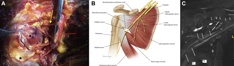
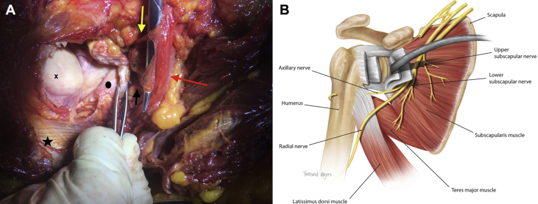
Successful healing of the subscapularis during anatomic total shoulder arthroplasty surgery is critical to optimize functional outcomes and avoid complications. The purpose of this study was to examine the upper and lower subscapularis nerve insertion in relation to the musculotendinous junction to estimate the risk of nerve injury. Our hypothesis was that arm position changes the risks to these nerves when exposing the anterior glenoid.
Twenty cadaveric shoulders were dissected, and the subscapular nerves were identified from the posterior cord of the brachial plexus to the muscle insertion. The nerve length from the origin to the muscle insertion and the distance to the myotendinous junction were measured in various shoulder positions including neutral, external, and internal rotation.
The mean length of the upper subscapular nerve was 51.4 ± 12.8 mm; that of the lower subscapular nerve was 50.5 ± 14 mm. The mean distance from the insertion of the upper subscapular nerve to the myotendinous junction 53.0 ± 14.7 mm with external rotation, 38.5 ± 9.7 mm with neutral rotation, and 30.0 ± 9.2 mm with internal rotation. The mean distance from the lower subscapular nerve to the myotendinous junction was 44.5 ± 13.8 mm with external rotation, 31.9 ± 9.3 mm with neutral rotation, and 25.4 ± 8.8 mm with internal rotation. The internally rotated position placed these nerves closest to the glenohumeral joint.
The upper and lower subscapular nerves insert in the muscle belly close to the myotendinous junction, putting them at risk of iatrogenic injury. Care must be taken to avoid damage with retractor placement in the anterior glenoid neck as these nerves are at risk of compression or torsional injury.
在解剖型全肩关节置换手术中,肩胛下肌的成功愈合对于优化功能结果和避免并发症至关重要。本研究的目的是检查肩胛下神经上下支与肌腱结合部的关系,以评估神经损伤的风险。我们的假设是,在暴露前方关节盂时,手臂位置会改变这些神经的风险。
解剖20具尸体肩部,从臂丛后束至肌肉附着点识别肩胛下神经。在包括中立位、外旋和内旋的各种肩部位置测量从神经起点到肌肉附着点的神经长度以及到肌腱结合部的距离。
肩胛上神经平均长度为51.4±12.8mm;肩胛下神经平均长度为50.5±14mm。肩胛上神经附着点到肌腱结合部的平均距离,外旋时为53.0±14.7mm,中立位时为38.5±9.7mm,内旋时为30.0±9.2mm。肩胛下神经到肌腱结合部的平均距离,外旋时为44.5±13.8mm,中立位时为31.9±9.3mm,内旋时为25.4±8.8mm。内旋位使这些神经最靠近盂肱关节。
肩胛下神经上下支附着于靠近肌腱结合部的肌腹,使其有发生医源性损伤的风险。在前方关节盂颈部放置牵开器时必须小心避免损伤,因为这些神经有受压或扭转损伤的风险。