Hendrickson Cole, Linden Katharina, Kreyer Stefan, Beilman Gregory, Scaravilli Vittorio, Wendorff Daniel, Necsoiu Corina, Batchinsky Andriy I, Cancio Leopoldo C, Chung Kevin K, Lusczek Elizabeth R
Department of Surgery, University of Minnesota, Minneapolis, MN 55455, USA.
US Army Institute of Surgical Research, Fort Sam Houston, TX 78234, USA.
Metabolites. 2019 Jul 12;9(7):142. doi: 10.3390/metabo9070142.
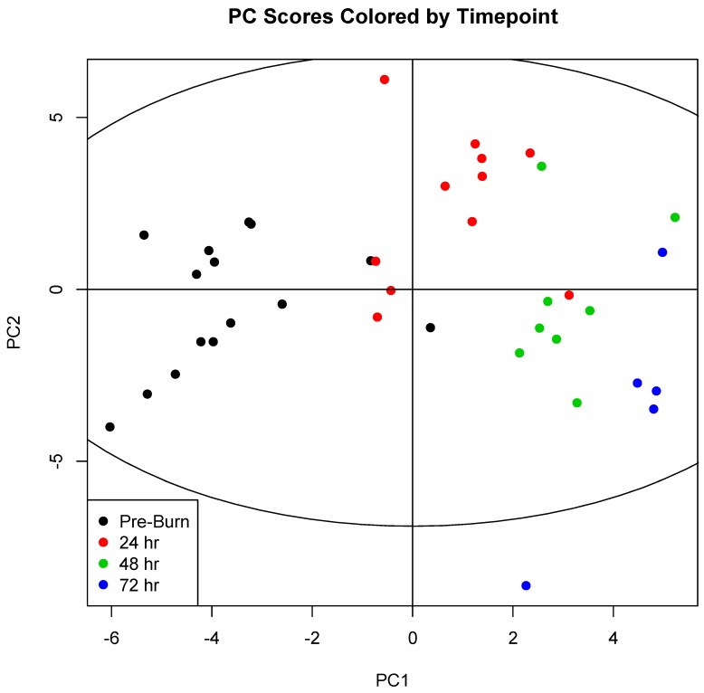
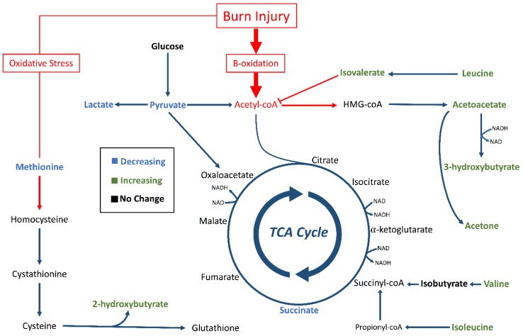
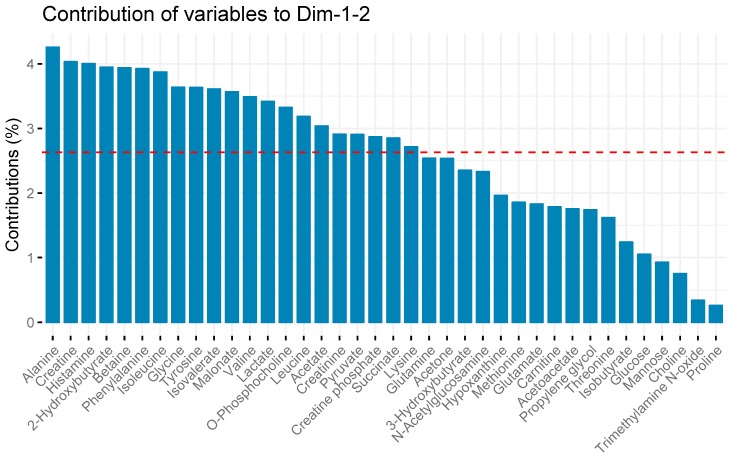
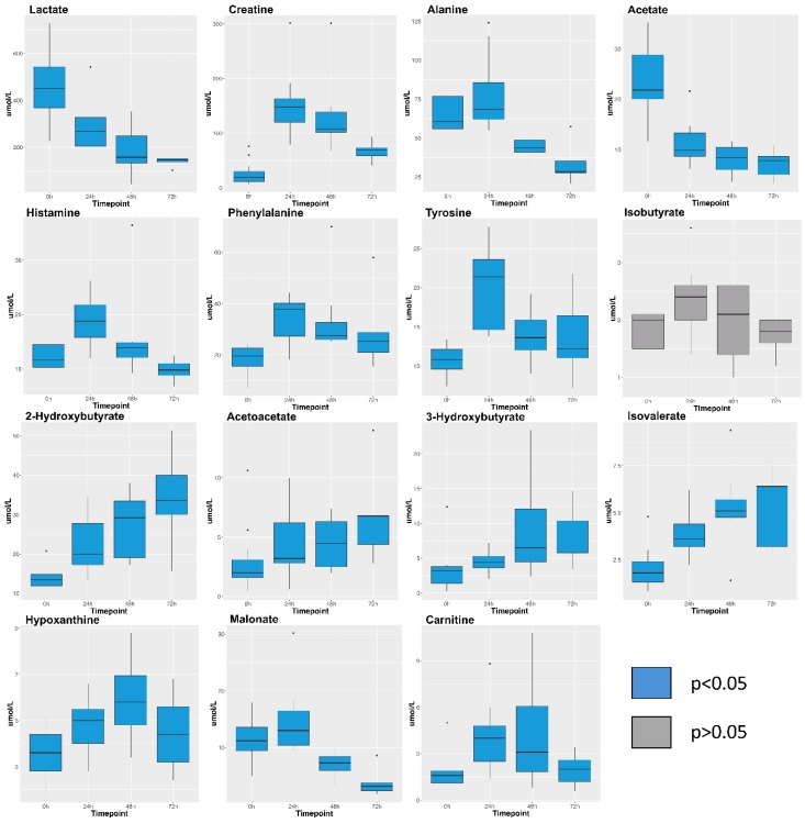
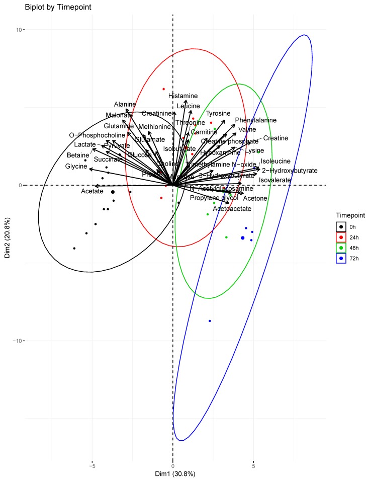
Burn injury initiates a hypermetabolic response leading to muscle catabolism and organ dysfunction but has not been well-characterized by high-throughput metabolomics. We examined changes in metabolism over the first 72 h post-burn using proton nuclear magnetic resonance (H-NMR) spectroscopy and serum from a porcine model of severe burn injury. We sought to quantify the changes in metabolism that occur over time in response to severe burn and smoke inhalation in this preliminary study. Fifteen pigs received 40% total body surface area (TBSA) burns with additional pine bark smoke inhalation. Arterial blood was drawn at baseline (pre-burn) and every 24 h until 72 h post-injury or death. The aqueous portion of each serum sample was analyzed using H-NMR spectroscopy and metabolite concentrations were used for principal component analysis (PCA). Thirty-eight metabolites were quantified in 39 samples. Of these, 31 showed significant concentration changes over time ( < 0.05). PCA revealed clustering of samples by time point on a 2D scores plot. The first 48 h post-burn were characterized by high concentrations of histamine, alanine, phenylalanine, and tyrosine. Later timepoints were characterized by rising concentrations of 2-hydroxybutyrate, 3-hydroxybutyrate, acetoacetate, and isovalerate. No significant differences in metabolism related to mortality were observed. Our work highlights the accumulation of organic acids resulting from fatty acid catabolism and oxidative stress. Further studies will be required to relate accumulation of the four organic carboxylates identified in this analysis to outcomes from burn injury.
烧伤会引发高代谢反应,导致肌肉分解代谢和器官功能障碍,但高通量代谢组学尚未对其进行充分表征。我们使用质子核磁共振(H-NMR)光谱和来自严重烧伤猪模型的血清,研究了烧伤后最初72小时内的代谢变化。在这项初步研究中,我们试图量化因严重烧伤和烟雾吸入随时间发生的代谢变化。15头猪接受了40%体表面积(TBSA)的烧伤,并额外吸入松树皮烟雾。在基线(烧伤前)以及伤后每24小时直至72小时或死亡时采集动脉血。使用H-NMR光谱分析每个血清样本的水相部分,并将代谢物浓度用于主成分分析(PCA)。在39个样本中对38种代谢物进行了定量。其中,31种代谢物的浓度随时间有显著变化(<0.05)。PCA在二维得分图上显示样本按时间点聚类。烧伤后的前48小时以组胺(histamine)、丙氨酸(alanine)、苯丙氨酸(phenylalanine)和酪氨酸(tyrosine)的高浓度为特征。后期时间点的特征是2-羟基丁酸(2-hydroxybutyrate)、3-羟基丁酸(3-hydroxybutyrate)、乙酰乙酸(acetoacetate)和异戊酸(isovalerate)的浓度升高。未观察到与死亡率相关的代谢显著差异。我们的工作突出了脂肪酸分解代谢和氧化应激导致的有机酸积累。需要进一步研究将本分析中鉴定的四种有机羧酸盐的积累与烧伤结局联系起来。