Kwon Sae Min, Ko Yong, Bang Seong Sik
Departments of Neurosurgery, College of Medicine, Hanyang University, 17 Haengdang-dong, Seongdong-gu, 133-792, Seoul, Republic of Korea.
Department of Neurosurgery, Keimyung University School of Medicine, 1095 Dalgubeol-daero, Dalseo-gu, Daegu, 42601, Republic of Korea.
BMC Neurol. 2019 Jul 23;19(1):176. doi: 10.1186/s12883-019-1392-5.
Primary intraosseous meningioma is a subset of extradural meningioma that arises in the bone, and only a few cases have been reported to date.
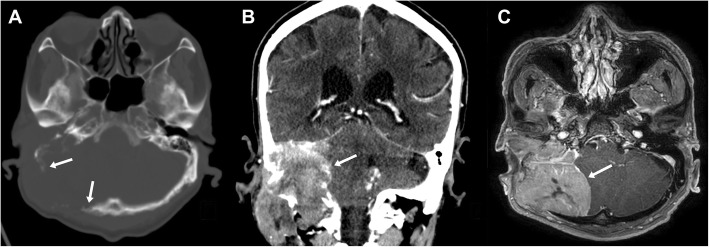
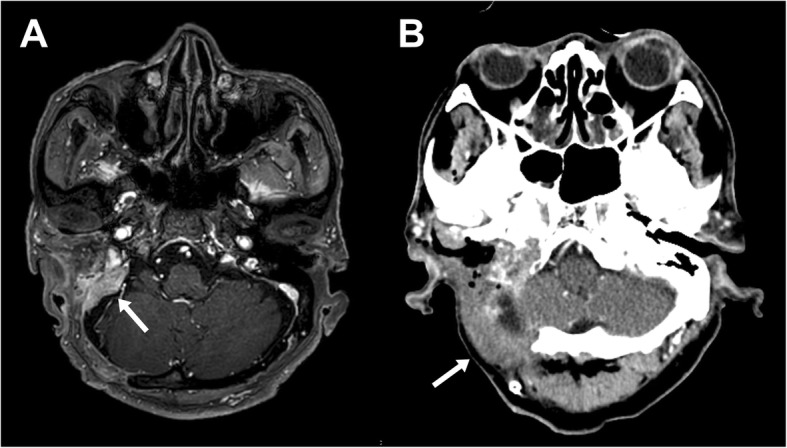
An 80-year-old man presented with decreased hearing on the right side accompanied by a disturbance of balance 10 months prior to admission. Magnetic resonance imaging revealed an 8 × 7 cm osteolytic mass in the right posterior fossa related to the petrous bone, with extension to the cervical region. During surgery, the tumor was found to be located extradurally, with no invasion of the dura. The tumor was removed entirely, apart from a small portion around the jugular foramen to avoid lower cranial nerve injury.
The final diagnosis was primary intraosseous osteolytic meningioma with atypical pathology. Here, we report a rare case of an osteolytic skull lesion in the skull base not invading the dura and with extensive bone destruction.
原发性骨内脑膜瘤是硬膜外脑膜瘤的一个亚型,起源于骨组织,迄今为止仅有少数病例报道。
一名80岁男性在入院前10个月出现右侧听力下降并伴有平衡障碍。磁共振成像显示右侧后颅窝与岩骨相关的一个8×7cm溶骨性肿块,延伸至颈部区域。手术中发现肿瘤位于硬膜外,未侵犯硬脑膜。除了颈静脉孔周围的一小部分以避免下颅神经损伤外,肿瘤被完全切除。
最终诊断为具有非典型病理的原发性骨内溶骨性脑膜瘤。在此,我们报告一例罕见的颅底溶骨性颅骨病变,未侵犯硬脑膜且伴有广泛骨质破坏。