Utsunomiya Yusuke, Mori Nobuyuki, Matsui Yuya, Katsushima Hiroki, Hashimoto Kenji, Furuta Akihiro
Department of Radiology, Osaka Red Cross Hospital, 5-30, Fudegasakicho, Tennoji-ku, Osaka 543-8555, Japan.
Department of Neurosurgery, Osaka Red Cross Hospital, Tennoji-ku, Osaka, Japan.
Radiol Case Rep. 2021 Aug 26;16(11):3300-3303. doi: 10.1016/j.radcr.2021.07.080. eCollection 2021 Nov.
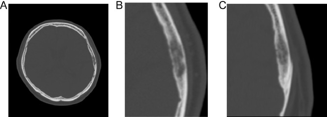
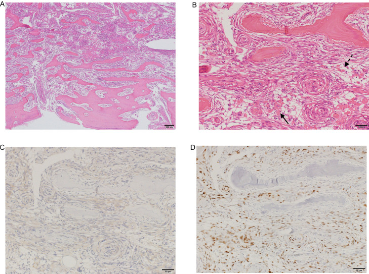
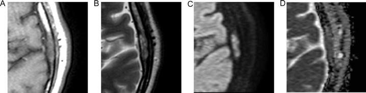
Metaplastic meningioma is a rare World Health Organization Grade I meningioma subtype, accounting for 0.2%-1.6% of all meningiomas. Primary extradural meningiomas represent less than 2% of all meningiomas, with intraosseous meningioma as a subtype of primary extradural meningiomas. Herein, we report the case of a 65-year-old male presenting with headache. His computed tomography scans showed an osteolytic left parietal bone mass, and magnetic resonance imaging revealed hyperintense dots in the mass on T1-weighted images. The mass was then resected and diagnosed on histopathological examination as an intraosseous metaplastic meningioma.
化生型脑膜瘤是一种罕见的世界卫生组织一级脑膜瘤亚型,占所有脑膜瘤的0.2%-1.6%。原发性硬膜外脑膜瘤占所有脑膜瘤的比例不到2%,骨内脑膜瘤是原发性硬膜外脑膜瘤的一种亚型。在此,我们报告一例65岁男性头痛病例。他的计算机断层扫描显示左侧顶骨有溶骨性肿块,磁共振成像显示该肿块在T1加权图像上有高信号点。随后该肿块被切除,经组织病理学检查诊断为骨内化生型脑膜瘤。