Internal Medicine, National Hospital Organization Nishi-Beppu Hospital, Tsurumi, Beppu, Oita, Japan.
Respiratory Medicine and Infectious Diseases, Oita University Faculty of Medicine, Idaigaoka, Hasama-machi, Yufu, Oita, Japan.
PLoS One. 2019 Jul 25;14(7):e0220346. doi: 10.1371/journal.pone.0220346. eCollection 2019.
Unusual radiological images may delay diagnosis of pulmonary tuberculosis. This study aimed to analyze the risk factors for an atypical radiological image in patients with pulmonary tuberculosis.
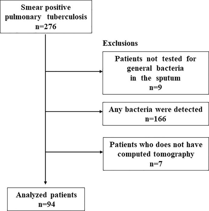
We retrospectively analyzed data from patients admitted to one hospital from January 2013 to December 2016 for sputum smear-positive lung tuberculosis who underwent chest computed tomography (CT) on admission. Patients whose sputum cultures were positive for general bacteria were excluded. Patient characteristics and laboratory data were compared between patients with cavity and those without and between patients with upper predominant lung involvement and those without.
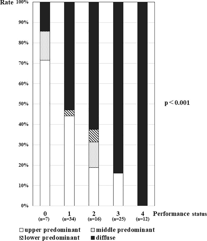
This study included 94 (93%) of 101 patients who underwent chest CT. The non-cavity group was older, had a greater number of females, had a lower C-reactive protein (CRP) level, and had a lower glomerular filtration rate. Multivariate analysis showed that a low CRP level (OR 0.808; 95% CI 0.674-0.967; p = 0.020) significantly predicted non-cavity pulmonary tuberculosis. The non-upper predominant lung involvement group was older and had a greater number of females, poorer performance status, a higher CRP level, and a lower serum albumin level. A poor performance status (OR 2.155; 95% CI 1.257-3.693; p = 0.005) was found to significantly predict pulmonary tuberculosis with non-upper predominant lung distributions.
A low CRP level and poor performance status were associated with non-cavity and non-upper predominant lung distribution, respectively, in patients with pulmonary tuberculosis. Tuberculosis patients with these characteristics may present unusual chest images.
不典型的影像学表现可能会延迟肺结核的诊断。本研究旨在分析肺结核患者影像学不典型的相关危险因素。
我们回顾性分析了 2013 年 1 月至 2016 年 12 月期间因痰涂片阳性肺结核而在一家医院住院的患者资料,所有患者入院时均接受了胸部计算机断层扫描(CT)检查。排除了痰普通细菌培养阳性的患者。比较了有空洞和无空洞患者、肺上叶病变为主和无肺上叶病变患者的患者特征和实验室数据。
本研究纳入了 101 例接受胸部 CT 检查的患者中的 94 例(93%)。非空洞组患者年龄较大,女性较多,C 反应蛋白(CRP)水平较低,肾小球滤过率较低。多因素分析显示,低 CRP 水平(OR 0.808;95% CI 0.674-0.967;p = 0.020)是预测非空洞性肺结核的重要因素。非肺上叶病变为主组患者年龄较大,女性较多,一般状况较差,CRP 水平较高,血清白蛋白水平较低。一般状况较差(OR 2.155;95% CI 1.257-3.693;p = 0.005)是预测肺上叶病变以外肺结核的重要因素。
在肺结核患者中,低 CRP 水平和一般状况较差分别与非空洞和非肺上叶病变分布相关。具有这些特征的肺结核患者可能会出现不典型的胸部影像。