• 文献检索
  • 文档翻译
  • 深度研究
  • 学术资讯
  • Suppr Zotero 插件Zotero 插件
  • 邀请有礼
  • 套餐&价格
  • 历史记录
应用&插件
Suppr Zotero 插件Zotero 插件浏览器插件Mac 客户端Windows 客户端微信小程序
定价
高级版会员购买积分包购买API积分包
服务
文献检索文档翻译深度研究API 文档MCP 服务
关于我们
关于 Suppr公司介绍联系我们用户协议隐私条款
关注我们

Suppr 超能文献

核心技术专利:CN118964589B侵权必究
粤ICP备2023148730 号-1Suppr @ 2026

文献检索

告别复杂PubMed语法,用中文像聊天一样搜索,搜遍4000万医学文献。AI智能推荐,让科研检索更轻松。

立即免费搜索

文件翻译

保留排版,准确专业,支持PDF/Word/PPT等文件格式,支持 12+语言互译。

免费翻译文档

深度研究

AI帮你快速写综述,25分钟生成高质量综述,智能提取关键信息,辅助科研写作。

立即免费体验

超声检查与后续髋关节发育不良X线检查结果之间的相关性。

Correlations between ultrasonographic and subsequent radiographic findings of developmental dysplasia of the hips.

作者信息

Tan Si Heng Sharon, Wu Cheng Han, Wong Keng Lin, Hui James Hoipo

机构信息

Department of Orthopaedic Surgery, University Orthopaedic, Hand and Reconstructive Microsurgery Cluster, National University Health System (NUHS), Singapore.

出版信息

Ultrasonography. 2019 Oct;38(4):43-51. doi: 10.14366/usg.18064. Epub 2019 Mar 16.

DOI:10.14366/usg.18064
PMID:31344996
原文链接:https://pmc.ncbi.nlm.nih.gov/articles/PMC6920622/
Abstract

PURPOSE

The study aimed to investigate the utility of ultrasonographic findings in predicting the subsequent radiographic parameters of developmental dysplasia of the hips.

METHODS

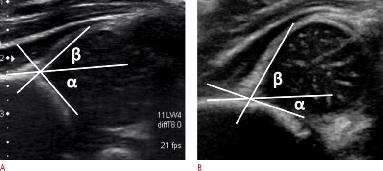
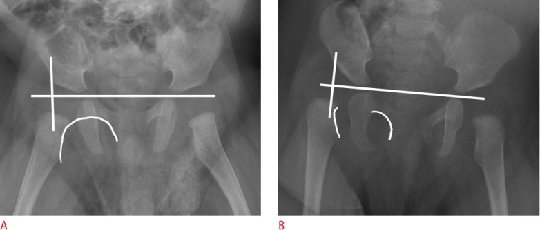
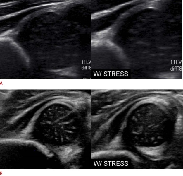
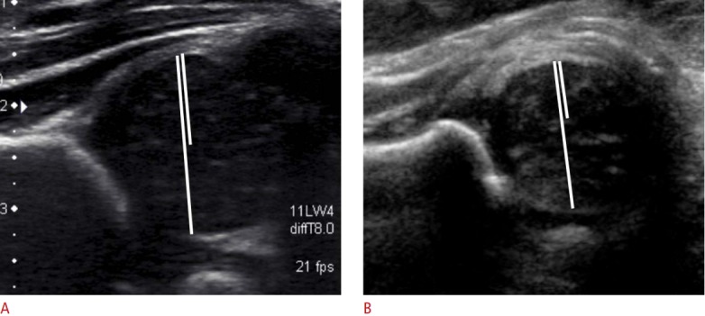
In this 12-year retrospective cohort study, all new-born infants with a positive clinical examination or risk factors were included. They were scheduled for hip ultrasonography in the first 3 months, and subsequent radiographs at 1 year of life. The ultrasonographic images were evaluated using the Graf classification, Harcke's dynamic screening method, and Terjesen's femoral head coverage method. The radiographic images were evaluated using the acetabular index and femoral head position. The overall ultrasonographic or radiographic findings were considered abnormal if they were classified as abnormal for any of their respective parameters. The overall ultrasonographic and radiographic parameters were correlated.

RESULTS

A total of 160 patients were included. The overall ultrasonographic and radiographic parameters showed no statistically significant difference (P=0.050). The sensitivity, specificity, and accuracy of the overall ultrasonographic parameters were 57.1%, 84.9%, and 81.3%, respectively. All three individual ultrasonographic parameters showed close correlations, with no statistically significant differences, with the overall radiographic findings and acetabular index (P>0.050). However, they showed a poor correlation, with a statistically significant difference, with the position of the femoral head (P<0.001), with the ultrasonographic parameters having an excellent negative predictive value of 100.0% for identifying an abnormal femoral head position.

CONCLUSION

The current study suggests that ultrasonographic findings evaluated in the first 3 months of life have a close correlation with radiographic findings evaluated at 1 year of life. The ultrasonographic parameters showed an excellent negative predictive value for abnormal femoral head position on radiographs.

摘要

目的

本研究旨在探讨超声检查结果在预测发育性髋关节发育不良后续X线参数方面的效用。

方法

在这项为期12年的回顾性队列研究中,纳入了所有临床检查阳性或有危险因素的新生儿。他们在出生后的前3个月接受髋关节超声检查,并在1岁时接受后续X线检查。超声图像采用Graf分类法、Harcke动态筛查法和Terjesen股骨头覆盖率法进行评估。X线图像采用髋臼指数和股骨头位置进行评估。如果整体超声或X线检查结果在各自参数中被分类为异常,则认为其整体检查结果异常。对整体超声和X线参数进行相关性分析。

结果

共纳入160例患者。整体超声和X线参数无统计学显著差异(P = 0.050)。整体超声参数的敏感性、特异性和准确性分别为57.1%、84.9%和81.3%。所有三个单独的超声参数与整体X线检查结果和髋臼指数均显示出密切相关性,无统计学显著差异(P>0.050)。然而,它们与股骨头位置的相关性较差,有统计学显著差异(P<0.001),超声参数在识别异常股骨头位置方面具有100.0%的出色阴性预测价值。

结论

本研究表明,出生后前3个月评估的超声检查结果与1岁时评估的X线检查结果密切相关。超声参数在预测X线片上异常股骨头位置方面具有出色的阴性预测价值。

https://cdn.ncbi.nlm.nih.gov/pmc/blobs/cbeb/6920622/049bac09bc00/usg-18064f5.jpg
https://cdn.ncbi.nlm.nih.gov/pmc/blobs/cbeb/6920622/a8589bb82862/usg-18064f1.jpg
https://cdn.ncbi.nlm.nih.gov/pmc/blobs/cbeb/6920622/72b8e03bb4cd/usg-18064f2.jpg
https://cdn.ncbi.nlm.nih.gov/pmc/blobs/cbeb/6920622/2f69a654bda9/usg-18064f3.jpg
https://cdn.ncbi.nlm.nih.gov/pmc/blobs/cbeb/6920622/e51fa8637cfe/usg-18064f4.jpg
https://cdn.ncbi.nlm.nih.gov/pmc/blobs/cbeb/6920622/049bac09bc00/usg-18064f5.jpg

相似文献

1
Correlations between ultrasonographic and subsequent radiographic findings of developmental dysplasia of the hips.超声检查与后续髋关节发育不良X线检查结果之间的相关性。
Ultrasonography. 2019 Oct;38(4):43-51. doi: 10.14366/usg.18064. Epub 2019 Mar 16.
2
The earliest timing of ultrasound in screening for developmental dysplasia of the hips.超声筛查发育性髋关节发育不良的最早时机。
Ultrasonography. 2019 Oct;38(4):321-326. doi: 10.14366/usg.18075. Epub 2019 Mar 3.
3
Correlation between ultrasonic and radiographic imaging of developmental dysplasia of the hip.发育性髋关节发育不良的超声与放射影像学相关性
J Child Orthop. 2019 Apr 1;13(2):155-160. doi: 10.1302/1863-2548.13.180165.
4
A Possible New Radiographic Predictor of Progression of Osteoarthritis in Developmental Dysplasia of the Hip: The Center Gap.髋关节发育不良性骨关节炎进展的一种可能的新影像学预测指标:中央间隙。
Clin Orthop Relat Res. 2018 Nov;476(11):2157-2166. doi: 10.1097/CORR.0000000000000458.
5
Exploration of femoral head coverage in screening developmental dysplasia of the hip in infants.婴儿发育性髋关节发育不良筛查中股骨头覆盖率的探讨。
J Med Ultrason (2001). 2019 Jan;46(1):129-135. doi: 10.1007/s10396-018-0911-0. Epub 2018 Oct 16.
6
Mild or Borderline Hip Dysplasia: Are We Characterizing Hips With a Lateral Center-Edge Angle Between 18° and 25° Appropriately?轻度或边缘性髋关节发育不良:我们是否正确地描述了外侧中心边缘角在 18°至 25°之间的髋关节?
Am J Sports Med. 2019 Jan;47(1):112-122. doi: 10.1177/0363546518810731.
7
Ultrasonographic findings in hips with a positive Ortolani sign and their relationship to Pavlik harness failure.髋关节 Ortolani 征阳性的超声表现及其与帕夫利克吊带治疗失败的关系。
J Bone Joint Surg Am. 2010 Jan;92(1):113-20. doi: 10.2106/JBJS.H.01880.
8
Do Radiographic Parameters of Dysplasia Improve to Normal Ranges After Bernese Periacetabular Osteotomy?伯尔尼髋臼周围截骨术后发育不良的影像学参数会改善至正常范围吗?
Clin Orthop Relat Res. 2017 Apr;475(4):1120-1127. doi: 10.1007/s11999-016-5077-8.
9
Discrepancy between ultrasonographic and radiographic findings in patients treated for developmental dysplasia of the hip; hip maturation on ultrasonography may not be consistent with radiographic hip maturation.超声与影像学表现不一致的患者在发育性髋关节发育不良的治疗;髋关节成熟度的超声可能与影像学髋关节成熟度不一致。
J Pediatr Orthop B. 2020 May;29(3):228-234. doi: 10.1097/BPB.0000000000000654.
10
Radiographic Follow-up of DDH in Infants: Are X-rays Necessary After a Normalized Ultrasound?婴儿发育性髋关节发育不良的影像学随访:超声检查结果正常后是否还需要进行X线检查?
J Pediatr Orthop. 2015 Sep;35(6):551-5. doi: 10.1097/BPO.0000000000000326.

引用本文的文献

1
Association Between Alpha Angle and Acetabular Index in Screening for Developmental Dysplasia of the Hip.阿尔法角与髋臼指数在髋关节发育不良筛查中的相关性
Cureus. 2024 Aug 6;16(8):e66334. doi: 10.7759/cureus.66334. eCollection 2024 Aug.
2
Association between early sonographic findings and acetabular index at the age of 6 months: a prospective observational study.6 月龄时超声早期表现与髋臼指数的相关性:一项前瞻性观察性研究。
BMC Pediatr. 2022 Apr 26;22(1):228. doi: 10.1186/s12887-022-03268-4.
3
Examining the Short-Term Natural History of Developmental Dysplasia of the Hip in Infancy: A Systematic Review.

本文引用的文献

1
Incorporating risk factors in the development of the screening programme for developmental dysplasia of the hips.
J Pediatr Orthop B. 2019 Mar;28(2):111-114. doi: 10.1097/BPB.0000000000000567.
2
Results of a universal ultrasonographic hip screening program at a single institution.单一机构的一项通用超声髋关节筛查项目的结果。
Acta Orthop Traumatol Turc. 2016;50(1):42-8. doi: 10.3944/AOTT.2016.15.0024.
3
The most relevant diagnostic criteria for developmental dysplasia of the hip: a study of British specialists.发育性髋关节发育不良最相关的诊断标准:一项对英国专家的研究。
婴幼儿期发育性髋关节发育不良的短期自然史研究:一项系统评价
Indian J Orthop. 2021 Sep 13;55(6):1372-1387. doi: 10.1007/s43465-021-00510-6. eCollection 2020 Dec.
4
Diagnostic Accuracy of Ultrasonography Method of Graf in the detection of Developmental Dysplasia of the Hip: A Meta-Analysis and Systematic Review.格拉夫超声检查法在检测发育性髋关节发育不良中的诊断准确性:一项荟萃分析与系统评价
Arch Bone Jt Surg. 2021 May;9(3):297-305. doi: 10.22038/abjs.2021.55292.2755.
BMC Musculoskelet Disord. 2016 Jan 19;17:38. doi: 10.1186/s12891-016-0867-4.
4
Do we need to follow up an early normal ultrasound with a later plain radiograph in children with a family history of developmental dysplasia of the hip?对于有髋关节发育不良家族史的儿童,我们是否需要在早期超声检查结果正常后再进行后期的X线平片检查?
Eur J Orthop Surg Traumatol. 2015 Oct;25(7):1171-5. doi: 10.1007/s00590-015-1668-4. Epub 2015 Jul 15.
5
Radiographic Follow-up of DDH in Infants: Are X-rays Necessary After a Normalized Ultrasound?婴儿发育性髋关节发育不良的影像学随访:超声检查结果正常后是否还需要进行X线检查?
J Pediatr Orthop. 2015 Sep;35(6):551-5. doi: 10.1097/BPO.0000000000000326.
6
Use of ultrasonography in developmental dysplasia of the hip.超声检查在发育性髋关节发育不良中的应用。
J Child Orthop. 2014 Mar;8(2):105-13. doi: 10.1007/s11832-014-0561-8. Epub 2014 Feb 9.
7
Selective ultrasound screening for developmental hip dysplasia: effect on management and late detected cases. A prospective survey during 1991-2006.发育性髋关节发育不良的选择性超声筛查:对治疗及晚期发现病例的影响。1991年至2006年的一项前瞻性调查。
Pediatr Radiol. 2014 Apr;44(4):410-24. doi: 10.1007/s00247-013-2838-3. Epub 2013 Dec 13.
8
Developmental dysplasia of the hip: controversies and current concepts.发育性髋关节发育不良:争议与当前概念
J Paediatr Child Health. 2012 Nov;48(11):963-72; quiz 972-3. doi: 10.1111/j.1440-1754.2012.02601.x.
9
Risk factors for developmental dysplasia of the hip: a meta-analysis.发育性髋关节发育不良的危险因素:荟萃分析。
Eur J Obstet Gynecol Reprod Biol. 2012 Nov;165(1):8-17. doi: 10.1016/j.ejogrb.2012.06.030. Epub 2012 Jul 21.
10
Reliability of ultrasonographic measurements in suspected patients of developmental dysplasia of the hip and correlation with the acetabular index.疑似发育性髋关节发育不良患者超声测量的可靠性及其与髋臼指数的相关性
Indian J Orthop. 2011 Nov;45(6):553-7. doi: 10.4103/0019-5413.87131.