• 文献检索
  • 文档翻译
  • 深度研究
  • 学术资讯
  • Suppr Zotero 插件Zotero 插件
  • 邀请有礼
  • 套餐&价格
  • 历史记录
应用&插件
Suppr Zotero 插件Zotero 插件浏览器插件Mac 客户端Windows 客户端微信小程序
定价
高级版会员购买积分包购买API积分包
服务
文献检索文档翻译深度研究API 文档MCP 服务
关于我们
关于 Suppr公司介绍联系我们用户协议隐私条款
关注我们

Suppr 超能文献

核心技术专利:CN118964589B侵权必究
粤ICP备2023148730 号-1Suppr @ 2026

文献检索

告别复杂PubMed语法,用中文像聊天一样搜索,搜遍4000万医学文献。AI智能推荐,让科研检索更轻松。

立即免费搜索

文件翻译

保留排版,准确专业,支持PDF/Word/PPT等文件格式,支持 12+语言互译。

免费翻译文档

深度研究

AI帮你快速写综述,25分钟生成高质量综述,智能提取关键信息,辅助科研写作。

立即免费体验

传统解剖标志是否有助于将 REBOA 导管尖端放置在最佳位置?一项非对照比较研究。

Does the conventional landmark help to place the tip of REBOA catheter in the optimal position? A non-controlled comparison study.

机构信息

1Advanced Critical Care and Emergency Center, Yokohama City University Medical Center, 4-57 Urafunecho, Minami-ku, Yokohama, Kanagawa 232-0024 Japan.

2Department of Emergency Medicine, Graduate School of Medicine, Yokohama City University, 3-9 Fukuura, Kanazawa-ku, Yokohama, Kanagawa 236-0004 Japan.

出版信息

World J Emerg Surg. 2019 Jul 16;14:35. doi: 10.1186/s13017-019-0255-0. eCollection 2019.

DOI:10.1186/s13017-019-0255-0
PMID:31346347
原文链接:https://pmc.ncbi.nlm.nih.gov/articles/PMC6635992/
Abstract

BACKGROUND

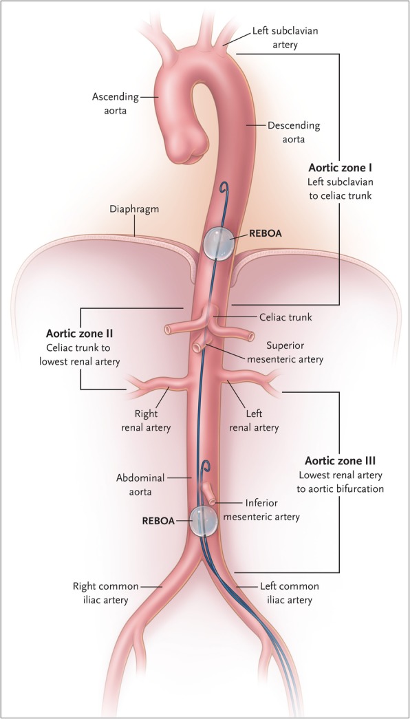
Resuscitative endovascular balloon occlusion of the aorta (REBOA) for patients with traumatic torso hemorrhagic shock is available to keep a minimum level of circulatory status as a bridge to definitive therapy. However, the trajectory for placement of REBOA in the aorta has not yet been clearly defined.

METHODS

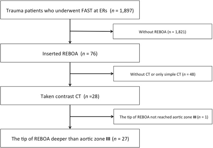
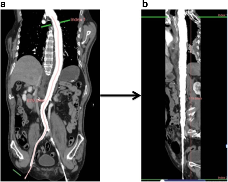
We conducted a retrospective observational cohort study in the two tertiary critical care and emergency center from December 2014 to October 2018. A total of 28 patients who underwent focused assessment with sonography for trauma (FAST) were studied via contrast computed tomography (CT), and 27 were analyzed.

RESULTS

We divided patients into two groups based on our CT findings. The REBOA deflate group included 16 patients, and the inflate group included 11 patients. The median trace value (interquartile range) of the blood vessel center line from the common femoral artery to the tip of REBOA (blood vessel length) and the length of REBOA itself from the common femoral artery to the tip of REBOA (REBOA insertion length) were 56.2 cm (54.5-57.2) and 55.2 cm (54.2-55.6), respectively ( < 0.0001) for the deflated group, and 51.4 cm (42.1-56.6) and 50.3 cm (42.3-55.0) ( = 0.594), respectively, for the inflated group.

CONCLUSIONS

If REBOA was deflated, it was placed 1.0 cm longer than the insertion length of REBOA catheter itself, but that was not the case when inflating REBOA. The individual difference was large to the extent that the balloon inflated and the extent to which the balloon was pushed back toward the caudal depending on the degree of blood pressure. Further studies would be needed to validate the study findings.

摘要

背景

主动脉球囊阻断复苏术(REBOA)用于创伤性躯干出血性休克患者,可保持最低循环状态,作为确定性治疗的桥梁。然而,主动脉内 REBOA 的放置轨迹尚未明确界定。

方法

我们对 2014 年 12 月至 2018 年 10 月期间在两个三级重症监护和急救中心进行了一项回顾性观察队列研究。对 28 例接受创伤超声重点评估(FAST)的患者进行了对比 CT 检查,其中 27 例进行了分析。

结果

根据我们的 CT 结果,我们将患者分为两组。REBOA 放气组包括 16 例患者,充气组包括 11 例患者。从股总动脉到 REBOA 尖端的血管中心线(血管长度)和从股总动脉到 REBOA 尖端的 REBOA 自身长度(REBOA 插入长度)的中位数(四分位间距)分别为 56.2cm(54.5-57.2)和 55.2cm(54.2-55.6)( < 0.0001),对于放气组,分别为 51.4cm(42.1-56.6)和 50.3cm(42.3-55.0)( = 0.594),分别为充气组。

结论

如果 REBOA 放气,它比 REBOA 导管自身插入长度长 1.0cm,但充气时并非如此。个体差异很大,程度取决于血压,气囊充气和气囊向尾部回推的程度。需要进一步的研究来验证研究结果。

https://cdn.ncbi.nlm.nih.gov/pmc/blobs/8315/6635992/de3ce41b2f03/13017_2019_255_Fig3_HTML.jpg
https://cdn.ncbi.nlm.nih.gov/pmc/blobs/8315/6635992/cc85a4832018/13017_2019_255_Fig1_HTML.jpg
https://cdn.ncbi.nlm.nih.gov/pmc/blobs/8315/6635992/abf1eda58980/13017_2019_255_Fig2_HTML.jpg
https://cdn.ncbi.nlm.nih.gov/pmc/blobs/8315/6635992/de3ce41b2f03/13017_2019_255_Fig3_HTML.jpg
https://cdn.ncbi.nlm.nih.gov/pmc/blobs/8315/6635992/cc85a4832018/13017_2019_255_Fig1_HTML.jpg
https://cdn.ncbi.nlm.nih.gov/pmc/blobs/8315/6635992/abf1eda58980/13017_2019_255_Fig2_HTML.jpg
https://cdn.ncbi.nlm.nih.gov/pmc/blobs/8315/6635992/de3ce41b2f03/13017_2019_255_Fig3_HTML.jpg

相似文献

1
Does the conventional landmark help to place the tip of REBOA catheter in the optimal position? A non-controlled comparison study.传统解剖标志是否有助于将 REBOA 导管尖端放置在最佳位置?一项非对照比较研究。
World J Emerg Surg. 2019 Jul 16;14:35. doi: 10.1186/s13017-019-0255-0. eCollection 2019.
2
Ultrasound-Guided Resuscitative Endovascular Balloon Occlusion of the Aorta in the Resuscitation Area.复苏区域内超声引导下的主动脉复苏性血管内球囊阻断术
J Emerg Med. 2017 May;52(5):715-722. doi: 10.1016/j.jemermed.2017.01.014. Epub 2017 Feb 17.
3
Computed tomography correlation of skeletal landmarks and vascular anatomy in civilian adult trauma patients: Implications for resuscitative endovascular balloon occlusion of the aorta.民用成年创伤患者骨骼标志和血管解剖的计算机断层扫描相关性:对主动脉复苏性血管内球囊阻断的影响。
J Trauma Acute Care Surg. 2019 Jul;87(1S Suppl 1):S138-S145. doi: 10.1097/TA.0000000000002247.
4
Anatomical landmarks for safely implementing resuscitative balloon occlusion of the aorta (REBOA) in zone 1 without fluoroscopy.在无荧光透视情况下安全实施1区主动脉复苏性球囊阻断术(REBOA)的解剖学标志。
Scand J Trauma Resusc Emerg Med. 2017 Jul 3;25(1):63. doi: 10.1186/s13049-017-0411-z.
5
Effect of resuscitative endovascular balloon occlusion of the aorta in hemodynamically unstable patients with multiple severe torso trauma: a retrospective study.主动脉球囊阻断复苏在血流动力学不稳定的多发严重躯干创伤患者中的作用:一项回顾性研究。
World J Emerg Surg. 2018 Oct 25;13:49. doi: 10.1186/s13017-018-0210-5. eCollection 2018.
6
Resuscitative Endovascular Balloon Occlusion of the Aorta Using a Low-Profile Device is Easy and Safe for Emergency Physicians in Cases of Life-Threatening Hemorrhage.使用低轮廓装置进行主动脉复苏性血管内球囊阻断术,对于面临危及生命出血情况的急诊医生来说既简便又安全。
J Emerg Med. 2018 Apr;54(4):410-418. doi: 10.1016/j.jemermed.2017.12.044. Epub 2018 Feb 10.
7
Resuscitative endovascular balloon occlusion of the aorta for control of noncompressible truncal hemorrhage in the abdomen and pelvis.用于控制腹部和盆腔不可压迫性躯干出血的主动脉复苏性血管内球囊阻断术。
Am J Surg. 2016 Dec;212(6):1222-1230. doi: 10.1016/j.amjsurg.2016.09.027. Epub 2016 Sep 30.
8
Resuscitative endovascular balloon occlusion of the aorta versus aortic cross clamping among patients with critical trauma: a nationwide cohort study in Japan.严重创伤患者中,主动脉内复苏性球囊阻断术与主动脉交叉钳夹术的比较:日本一项全国性队列研究
Crit Care. 2016 Dec 15;20(1):400. doi: 10.1186/s13054-016-1577-x.
9
Evaluation of the safety and feasibility of resuscitative endovascular balloon occlusion of the aorta.主动脉复苏性血管内球囊阻断术的安全性和可行性评估。
J Trauma Acute Care Surg. 2015 May;78(5):897-903; discussion 904. doi: 10.1097/TA.0000000000000614.
10
A new method using surface landmarks to locate resuscitative endovascular balloon occlusion of the aorta based on a retrospective CTA study.基于一项回顾性CTA研究,一种利用体表标志定位主动脉复苏性血管内球囊阻断术的新方法。
Eur J Trauma Emerg Surg. 2022 Jun;48(3):1945-1953. doi: 10.1007/s00068-021-01686-0. Epub 2021 May 21.

引用本文的文献

1
Age affects vascular morphology and predictiveness of anatomical landmarks for aortic zones in trauma patients: implications for resuscitative endovascular balloon occlusion of the aorta.年龄影响创伤患者主动脉区域的血管形态及解剖标志的预测性:对主动脉复苏性血管内球囊阻断术的启示。
Eur J Trauma Emerg Surg. 2024 Dec;50(6):3099-3108. doi: 10.1007/s00068-024-02512-z. Epub 2024 Apr 24.

本文引用的文献

1
Initial Care of the Severely Injured Patient.重伤患者的初始护理。
N Engl J Med. 2019 Feb 21;380(8):763-770. doi: 10.1056/NEJMra1609326.
2
Guidelines for a system-wide multidisciplinary approach to institutional resuscitative endovascular balloon occlusion of the aorta implementation.系统范围多学科方法实施机构复苏性主动脉内球囊阻断指南。
J Trauma Acute Care Surg. 2019 Feb;86(2):337-343. doi: 10.1097/TA.0000000000002138.
3
CE: The Use of Resuscitative Endovascular Balloon Occlusion of the Aorta in Treating Hemorrhagic Shock from Severe Trauma.
CE:主动脉内复苏性球囊阻断术在治疗严重创伤性出血性休克中的应用。
Am J Nurs. 2018 Oct;118(10):22-28. doi: 10.1097/01.NAJ.0000546376.73926.5b.
4
Radiofrequency Identification of the ER-REBOA: Confirmation of Placement Without Fluoroscopy.急诊室可重复使用球囊阻断主动脉术的射频识别:无需透视确认放置位置
Mil Med. 2019 Mar 1;184(3-4):e285-e289. doi: 10.1093/milmed/usy187.
5
Resuscitative endovascular balloon occlusion of the aorta for non-traumatic intra-abdominal hemorrhage.主动脉内球囊阻断复苏术治疗非创伤性腹腔内出血。
Eur J Trauma Emerg Surg. 2019 Aug;45(4):713-718. doi: 10.1007/s00068-018-0973-0. Epub 2018 Jun 19.
6
A systematic review and meta-analysis of the use of resuscitative endovascular balloon occlusion of the aorta in the management of major exsanguination.一项关于在严重失血管理中使用主动脉复苏性血管内球囊阻断术的系统评价和荟萃分析。
Eur J Trauma Emerg Surg. 2018 Aug;44(4):535-550. doi: 10.1007/s00068-018-0959-y. Epub 2018 May 21.
7
Resuscitative endovascular balloon occlusion of the aorta performed by emergency physicians for traumatic hemorrhagic shock: a case series from Japanese emergency rooms.急诊医生行主动脉腔内球囊阻断复苏治疗创伤性失血性休克:日本急诊室的病例系列。
Crit Care. 2018 Apr 21;22(1):103. doi: 10.1186/s13054-018-2032-y.
8
Use of Resuscitative Endovascular Balloon Occlusion of the Aorta for Proximal Aortic Control in Patients With Severe Hemorrhage and Arrest.使用主动脉抢救性血管内球囊阻断术控制严重出血性休克患者的近端主动脉。
JAMA Surg. 2018 Feb 1;153(2):130-135. doi: 10.1001/jamasurg.2017.3549.
9
Resuscitative Endovascular Balloon Occlusion of the Aorta in trauma: a systematic review of the literature.创伤性主动脉腔内球囊阻断复苏:文献系统评价。
World J Emerg Surg. 2017 Aug 29;12:42. doi: 10.1186/s13017-017-0153-2. eCollection 2017.
10
Anatomical landmarks for safely implementing resuscitative balloon occlusion of the aorta (REBOA) in zone 1 without fluoroscopy.在无荧光透视情况下安全实施1区主动脉复苏性球囊阻断术(REBOA)的解剖学标志。
Scand J Trauma Resusc Emerg Med. 2017 Jul 3;25(1):63. doi: 10.1186/s13049-017-0411-z.