• 文献检索
  • 文档翻译
  • 深度研究
  • 学术资讯
  • Suppr Zotero 插件Zotero 插件
  • 邀请有礼
  • 套餐&价格
  • 历史记录
应用&插件
Suppr Zotero 插件Zotero 插件浏览器插件Mac 客户端Windows 客户端微信小程序
定价
高级版会员购买积分包购买API积分包
服务
文献检索文档翻译深度研究API 文档MCP 服务
关于我们
关于 Suppr公司介绍联系我们用户协议隐私条款
关注我们

Suppr 超能文献

核心技术专利:CN118964589B侵权必究
粤ICP备2023148730 号-1Suppr @ 2026

文献检索

告别复杂PubMed语法,用中文像聊天一样搜索,搜遍4000万医学文献。AI智能推荐,让科研检索更轻松。

立即免费搜索

文件翻译

保留排版,准确专业,支持PDF/Word/PPT等文件格式,支持 12+语言互译。

免费翻译文档

深度研究

AI帮你快速写综述,25分钟生成高质量综述,智能提取关键信息,辅助科研写作。

立即免费体验

糖缀合胆汁酸通过隐窝裂变驱动多层腺体向初始化生腺形成,在小鼠模型中。

Glyco-conjugated bile acids drive the initial metaplastic gland formation from multi-layered glands through crypt-fission in a murine model.

机构信息

Center for Experimental and Molecular Medicine (CEMM), Academic Medical Center, Amsterdam, The Netherlands.

Tytgat Institute for Liver and Intestinal Research, Academic Medical Center, Amsterdam, The Netherlands.

出版信息

PLoS One. 2019 Jul 26;14(7):e0220050. doi: 10.1371/journal.pone.0220050. eCollection 2019.

DOI:10.1371/journal.pone.0220050
PMID:31348796
原文链接:https://pmc.ncbi.nlm.nih.gov/articles/PMC6660124/
Abstract

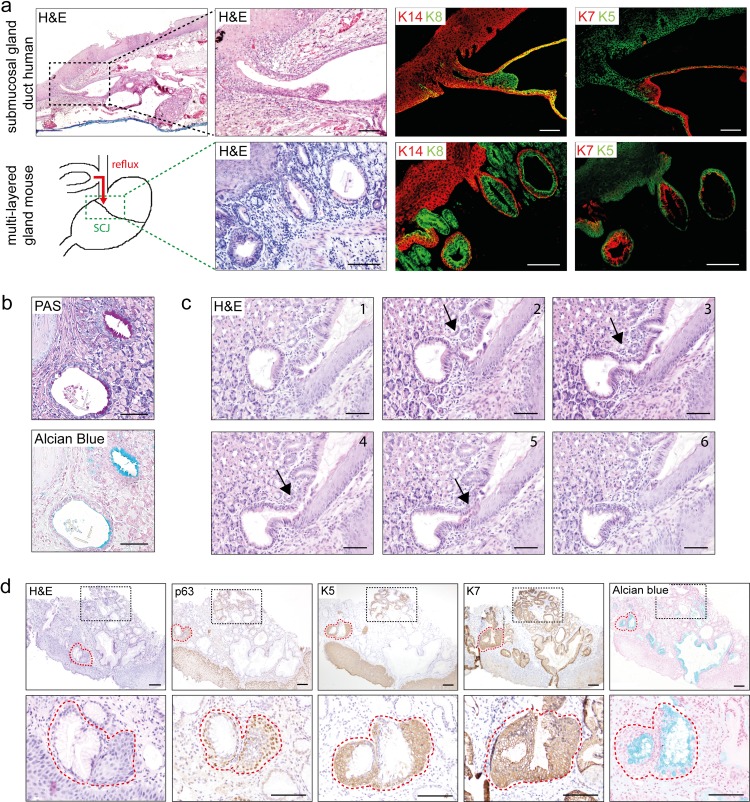
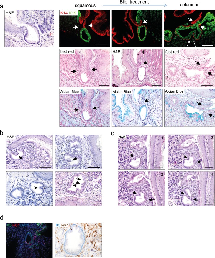
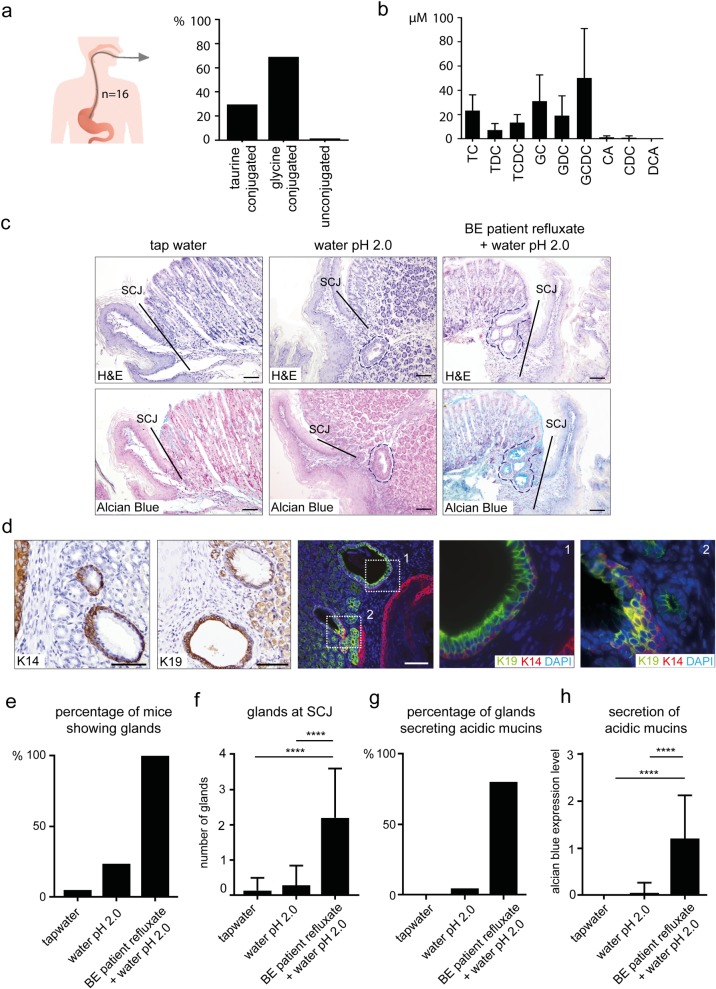
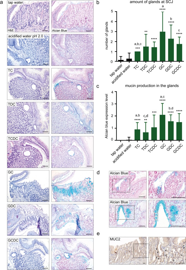
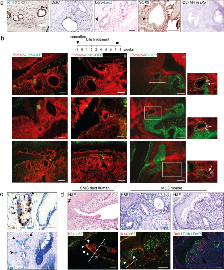
Bile acid reflux is known to be associated with the development of Barrett's esophagus and esophageal adenocarcinoma (EAC), yet the role of specific bile acids and the mechanism behind the metaplastic changes is unclear. Here, we demonstrate that multi-layered glandular structures at the squamo-columnar junction in mice contain multiple cell lineages, which resemble the human esophageal submucosal gland ducts. Exposing mice to patient's refluxates induced expansion of multi-layered glandular structures and development of columnar metaplasia at the squamo-columnar junction. The glycine conjugated bile acids induced an intestinal type of metaplasia more typical for Barrett's esophagus. Through lineage tracing, we excluded the involvement of K5+, DCLK1+, and LGR5+ progenitor cells as the primary source in the development of the glandular metaplastic epithelium. We show that the mechanism behind development of metaplasia involves crypt fission and may be independent of stem cell proliferation. Our findings support the hypothesis that in humans, BE arises from non-squamous cells residing in submucosal gland ducts and that induction of intestinal type of metaplasia is most effectively induced by glycine-conjugated bile acids. These novel insights may lead to more effective strategies to prevent development of Barrett's esophagus and esophageal adenocarcinoma.

摘要

胆汁酸反流已知与 Barrett 食管和食管腺癌(EAC)的发展有关,但特定胆汁酸的作用及其化生变化背后的机制尚不清楚。在这里,我们证明了小鼠鳞柱状交界处的多层腺状结构包含多种细胞谱系,类似于人类食管黏膜下腺导管。将患者的反流物暴露于小鼠中会诱导多层腺状结构的扩张,并在鳞柱状交界处发展出柱状化生。甘氨酸结合的胆汁酸诱导出更类似于 Barrett 食管的肠型化生。通过谱系追踪,我们排除了 K5+、DCLK1+和 LGR5+祖细胞作为腺性化生上皮发育的主要来源。我们表明,化生发展背后的机制涉及隐窝分裂,并且可能与干细胞增殖无关。我们的发现支持这样一种假说,即在人类中,BE 源自位于黏膜下腺导管中的非鳞状细胞,并且甘氨酸结合的胆汁酸最有效地诱导肠型化生。这些新的见解可能会导致更有效的策略来预防 Barrett 食管和食管腺癌的发展。

https://cdn.ncbi.nlm.nih.gov/pmc/blobs/dc22/6660124/4cda8c176fe2/pone.0220050.g005.jpg
https://cdn.ncbi.nlm.nih.gov/pmc/blobs/dc22/6660124/2613ce571074/pone.0220050.g001.jpg
https://cdn.ncbi.nlm.nih.gov/pmc/blobs/dc22/6660124/7f8652ab0ccc/pone.0220050.g002.jpg
https://cdn.ncbi.nlm.nih.gov/pmc/blobs/dc22/6660124/9e1bc1cf33a2/pone.0220050.g003.jpg
https://cdn.ncbi.nlm.nih.gov/pmc/blobs/dc22/6660124/94899922fb3d/pone.0220050.g004.jpg
https://cdn.ncbi.nlm.nih.gov/pmc/blobs/dc22/6660124/4cda8c176fe2/pone.0220050.g005.jpg

相似文献

1
Glyco-conjugated bile acids drive the initial metaplastic gland formation from multi-layered glands through crypt-fission in a murine model.糖缀合胆汁酸通过隐窝裂变驱动多层腺体向初始化生腺形成,在小鼠模型中。
PLoS One. 2019 Jul 26;14(7):e0220050. doi: 10.1371/journal.pone.0220050. eCollection 2019.
2
Origins of Metaplasia in Barrett's Esophagus: Is this an Esophageal Stem or Progenitor Cell Disease?巴雷特食管化生的起源:这是一种食管干细胞或祖细胞疾病吗?
Dig Dis Sci. 2018 Aug;63(8):2005-2012. doi: 10.1007/s10620-018-5069-5.
3
Acidic Bile Salts Induce Epithelial to Mesenchymal Transition via VEGF Signaling in Non-Neoplastic Barrett's Cells.酸性胆汁盐通过 VEGF 信号诱导非肿瘤性 Barrett 细胞上皮间质转化。
Gastroenterology. 2019 Jan;156(1):130-144.e10. doi: 10.1053/j.gastro.2018.09.046. Epub 2018 Sep 27.
4
Expression of the putative stem cell marker Musashi-1 in Barrett's esophagus and esophageal adenocarcinoma.Musashi-1 作为一种假定的干细胞标志物在 Barrett 食管和食管腺癌中的表达。
Dis Esophagus. 2010 Sep;23(7):580-9. doi: 10.1111/j.1442-2050.2010.01061.x. Epub 2010 Apr 29.
5
From Reflux Esophagitis to Esophageal Adenocarcinoma.从反流性食管炎到食管腺癌
Dig Dis. 2016;34(5):483-90. doi: 10.1159/000445225. Epub 2016 Jun 22.
6
Transitional basal cells at the squamous-columnar junction generate Barrett's oesophagus.鳞状柱状交界处的移行性基底细胞会引发巴雷特食管。
Nature. 2017 Oct 26;550(7677):529-533. doi: 10.1038/nature24269. Epub 2017 Oct 12.
7
Acid, bile, and CDX: the ABCs of making Barrett's metaplasia.酸、胆汁与CDX:形成巴雷特化生的基本要素
Am J Physiol Gastrointest Liver Physiol. 2008 Aug;295(2):G211-8. doi: 10.1152/ajpgi.90250.2008. Epub 2008 Jun 12.
8
The role of bile acids in the neoplastic progression of Barrett's esophagus - a short representative overview.胆汁酸在巴雷特食管肿瘤进展中的作用——简短代表性综述。
Z Gastroenterol. 2012 Sep;50(9):1028-34. doi: 10.1055/s-0032-1312922. Epub 2012 Sep 10.
9
Individual crypt genetic heterogeneity and the origin of metaplastic glandular epithelium in human Barrett's oesophagus.人类巴雷特食管中单个隐窝的遗传异质性及化生腺上皮的起源
Gut. 2008 Aug;57(8):1041-8. doi: 10.1136/gut.2007.143339. Epub 2008 Feb 27.
10
Transcommitment: Paving the Way to Barrett's Metaplasia.转分化:通向巴雷特化生之路的铺垫。
Adv Exp Med Biol. 2016;908:183-212. doi: 10.1007/978-3-319-41388-4_10.

引用本文的文献

1
Increased PXR and Suppressed T-Cell Signaling Are Associated With Malignant Degeneration of Barrett's Esophagus.孕烷X受体增加和T细胞信号传导受抑制与巴雷特食管的恶性变相关。
Gastro Hep Adv. 2022 Aug 24;2(1):63-71. doi: 10.1016/j.gastha.2022.08.005. eCollection 2023.
2
Inflammatory microenvironment in gastric premalignant lesions: implication and application.胃前病变中的炎症微环境:意义与应用。
Front Immunol. 2023 Nov 15;14:1297101. doi: 10.3389/fimmu.2023.1297101. eCollection 2023.
3
Molecular Biology and Clinical Management of Esophageal Adenocarcinoma.

本文引用的文献

1
TGF-β Family Signaling Pathways in Cellular Dormancy.细胞休眠中的转化生长因子-β家族信号通路
Trends Cancer. 2019 Jan;5(1):66-78. doi: 10.1016/j.trecan.2018.10.010. Epub 2018 Nov 28.
2
Esomeprazole and aspirin in Barrett's oesophagus (AspECT): a randomised factorial trial.埃索美拉唑和阿司匹林在 Barrett 食管(AspECT)中的应用:一项随机析因试验。
Lancet. 2018 Aug 4;392(10145):400-408. doi: 10.1016/S0140-6736(18)31388-6. Epub 2018 Jul 26.
3
Regenerative proliferation of differentiated cells by mTORC1-dependent paligenosis.mTORC1 依赖性返老还童促进分化细胞的再生增殖。
食管腺癌的分子生物学与临床管理
Cancers (Basel). 2023 Nov 14;15(22):5410. doi: 10.3390/cancers15225410.
4
Diagnostic Principles for Chronic Gastritis Associated with Duodenogastric Reflux.十二指肠-胃反流性慢性胃炎的诊断原则
Diagnostics (Basel). 2023 Jan 4;13(2):186. doi: 10.3390/diagnostics13020186.
5
Bile Acids: Physiological Activity and Perspectives of Using in Clinical and Laboratory Diagnostics.胆汁酸:生理活性及在临床与实验室诊断中的应用展望。
Molecules. 2022 Nov 13;27(22):7830. doi: 10.3390/molecules27227830.
6
Roles and action mechanisms of bile acid-induced gastric intestinal metaplasia: a review.胆汁酸诱导胃黏膜肠化生的作用及机制:综述
Cell Death Discov. 2022 Apr 4;8(1):158. doi: 10.1038/s41420-022-00962-1.
7
Inhibition of the BMP pathway prevents development of Barrett's-associated adenocarcinoma in a surgical rat model.抑制 BMP 通路可预防手术大鼠模型中 Barrett 相关腺癌的发生。
Dis Esophagus. 2022 May 10;35(5). doi: 10.1093/dote/doab072.
8
Impact of Deoxycholic Acid on Oesophageal Adenocarcinoma Invasion: Effect on Matrix Metalloproteinases.脱氧胆酸对食管腺癌侵袭的影响:对基质金属蛋白酶的影响。
Int J Mol Sci. 2020 Oct 28;21(21):8042. doi: 10.3390/ijms21218042.
EMBO J. 2018 Apr 3;37(7). doi: 10.15252/embj.201798311. Epub 2018 Feb 21.
4
Transitional basal cells at the squamous-columnar junction generate Barrett's oesophagus.鳞状柱状交界处的移行性基底细胞会引发巴雷特食管。
Nature. 2017 Oct 26;550(7677):529-533. doi: 10.1038/nature24269. Epub 2017 Oct 12.
5
Origin of Barrett's Epithelium: Esophageal Submucosal Glands.巴雷特食管上皮的起源:食管黏膜下腺。
Cell Mol Gastroenterol Hepatol. 2017 Mar 6;4(1):153-156. doi: 10.1016/j.jcmgh.2017.01.016. eCollection 2017 Jul.
6
Ductular and proliferative response of esophageal submucosal glands in a porcine model of esophageal injury and repair.猪食管损伤与修复模型中食管黏膜下腺的导管和增殖反应
Am J Physiol Gastrointest Liver Physiol. 2017 Sep 1;313(3):G180-G191. doi: 10.1152/ajpgi.00036.2017. Epub 2017 Jun 1.
7
The Barrett's Gland in Phenotype Space.表型空间中的巴雷特腺。
Cell Mol Gastroenterol Hepatol. 2014 Nov 12;1(1):41-54. doi: 10.1016/j.jcmgh.2014.10.001. eCollection 2015 Jan.
8
Proton Pump Inhibitors Do Not Reduce the Risk of Esophageal Adenocarcinoma in Patients with Barrett's Esophagus: A Systematic Review and Meta-Analysis.质子泵抑制剂不能降低巴雷特食管患者食管腺癌的风险:一项系统评价和荟萃分析。
PLoS One. 2017 Jan 10;12(1):e0169691. doi: 10.1371/journal.pone.0169691. eCollection 2017.
9
Paneth Cell-Rich Regions Separated by a Cluster of Lgr5+ Cells Initiate Crypt Fission in the Intestinal Stem Cell Niche.由一簇Lgr5 +细胞分隔的富含潘氏细胞的区域启动肠道干细胞龛中的隐窝裂变。
PLoS Biol. 2016 Jun 27;14(6):e1002491. doi: 10.1371/journal.pbio.1002491. eCollection 2016 Jun.
10
Intestinal Stem Cell Markers in the Intestinal Metaplasia of Stomach and Barrett's Esophagus.胃及巴雷特食管肠化生中的肠道干细胞标志物
PLoS One. 2015 May 21;10(5):e0127300. doi: 10.1371/journal.pone.0127300. eCollection 2015.