Department of Pathology, Ruijin Hospital, Shanghai Jiaotong University School of Medicine, No. 197 Ruijin Er Road, Huangpu District, Shanghai 200025, China.
Department of Pathology, Jinjiang People's Hospital, No. 28 Zhongzhou Road, Jingjiang, Jiangsu 214500, China.
Biomed Res Int. 2019 Jul 2;2019:5026860. doi: 10.1155/2019/5026860. eCollection 2019.
Calcifying fibrous tumor (CFT) is a very rare begin fibroblastic tumor featuring a widely anatomical distribution and may mimic various spindle cell tumors. Misdiagnosis and hence mistreatment are likely caused due to unfamiliarity to clinicians or junior pathologists. We collected a relatively large series of CFTs in our institution aiming at further summarizing their clinicopathologic features in Chinese patients and discussing the diagnosis and differential diagnosis in clinical practice.
Clinicopathologic data of 22 CFTs were retrospectively reviewed. Histologic features were reevaluated and summarized. Immunostaining markers include CD34, SMA, Desmin, keratin, S100, ALK1, CD117, IgG, IgG4, and Ki-67. Follow-up of all cases was performed.
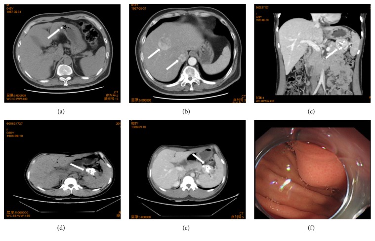
22 CFTs include gastric (n=8), pulmonary (n=2), hepatic (n=2), cervical (n=1), appendiceal (n=1), esophageal (n=1), retroperitoneal (n=1), intra-abdominal (n=1), diaphragmatic (n=1), spermatic cord and scrotum (n=1), anconeal (n=1), mesenteric (n=1), and omental (n=1) lesions. Coexisting hepatocellular carcinoma, pancreatic carcinoma, pheochromocytoma, Castleman disease, and leiomyoma of the uterus and other metabolic or functional disorders were also appreciated. CFT histologically features spindle cells embedded dense hyalinized stroma with scattered psammomatous calcifications and lymphoplasmacytic infiltration and immunohistochemically for CD34. None of any individuals die of CFT per se.
Our study discloses that CFT is a bona fide benign fibroblastic lesion, regardless of its developing location. Involvement of digestive tract seems much more common in the Chinese population. Awareness of the clinicopathologic characteristics of this rare entity and its mimickers contribute to avoiding misdiagnosis and mistreatment in clinical practice.
钙化性纤维瘤(CFT)是一种非常罕见的起始纤维母细胞瘤,具有广泛的解剖分布,可能模仿各种梭形细胞肿瘤。由于临床医生或初级病理学家不熟悉,误诊和因此的治疗不当很可能发生。我们在机构中收集了相对较大系列的 CFT,旨在进一步总结中国患者的临床病理特征,并讨论临床实践中的诊断和鉴别诊断。
回顾性分析 22 例 CFT 的临床病理资料。重新评估和总结组织学特征。免疫组织化学标志物包括 CD34、SMA、结蛋白、角蛋白、S100、ALK1、CD117、IgG、IgG4 和 Ki-67。对所有病例进行随访。
22 例 CFT 包括胃(n=8)、肺(n=2)、肝(n=2)、宫颈(n=1)、阑尾(n=1)、食管(n=1)、腹膜后(n=1)、腹腔内(n=1)、膈肌(n=1)、肌腱和阴囊(n=1)、肘(n=1)、肠系膜(n=1)和网膜(n=1)病变。还注意到共存的肝细胞癌、胰腺癌、嗜铬细胞瘤、Castleman 病、子宫平滑肌瘤和其他代谢或功能障碍。CFT 的组织学特征为嵌入致密玻璃样基质的梭形细胞,伴有散在的砂粒体钙化和淋巴浆细胞浸润,并免疫组织化学染色为 CD34。没有任何一个人因 CFT 本身而死亡。
我们的研究表明,CFT 是一种真正的良性纤维母细胞瘤病变,无论其发生部位如何。中国人群中似乎更常见累及消化道。了解这种罕见实体及其类似物的临床病理特征有助于避免临床实践中的误诊和治疗不当。