School of Physics and Astronomy University of Minnesota, Twin Cities Physics and Nanotechnology (PAN), 115 Union Street SE, Minneapolis, MN, 55455, USA.
Middlebury College, 14 Old Chapel Road, Middlebury, VT, 05753, USA.
Nat Commun. 2019 Jul 30;10(1):3400. doi: 10.1038/s41467-019-11384-6.
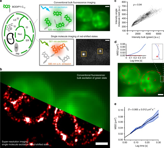
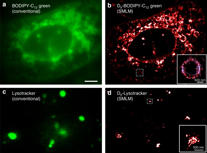
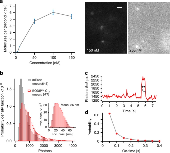
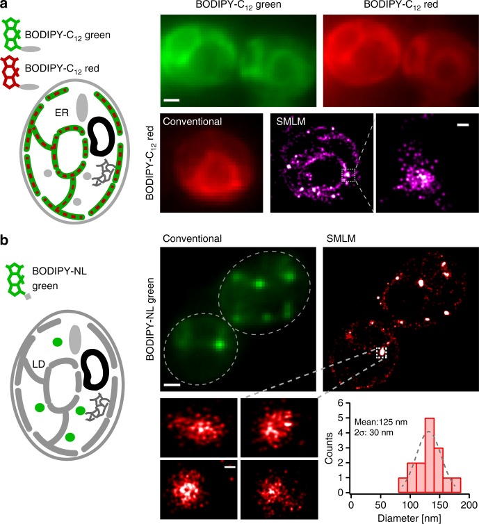
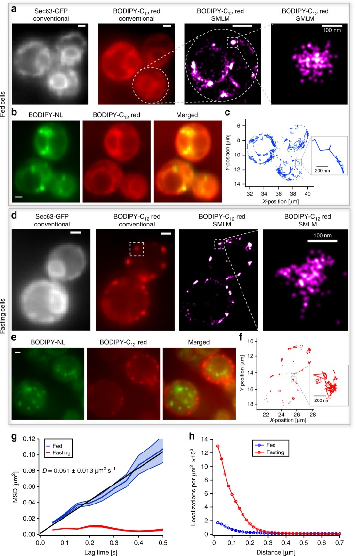
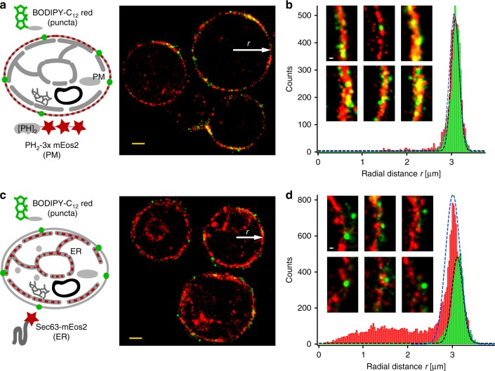
Single-molecule localization microscopy (SMLM) is a rapidly evolving technique to resolve subcellular structures and single-molecule dynamics at the nanoscale. Here, we employ conventional BODIPY conjugates for live-cell SMLM via their previously reported red-shifted ground-state dimers (D), which transiently form through bi-molecular encounters and emit bright single-molecule fluorescence. We employ the versatility of D-state SMLM to resolve the nanoscopic spatial regulation and dynamics of single fatty acid analogs (FAas) and lipid droplets (LDs) in living yeast and mammalian cells with two colors. In fed cells, FAas localize to the endoplasmic reticulum and LDs of ~125 nm diameter. Upon fasting, however, FAas form dense, non-LD clusters of ~100 nm diameter at the plasma membrane and transition from free diffusion to confined immobilization. Our reported SMLM capability of conventional BODIPY conjugates is further demonstrated by imaging lysosomes in mammalian cells and enables simple and versatile live-cell imaging of sub-cellular structures at the nanoscale.
单分子定位显微镜(SMLM)是一种快速发展的技术,可在纳米尺度上解析亚细胞结构和单分子动力学。在这里,我们通过先前报道的红移基态二聚体(D)将常规 BODIPY 缀合物用于活细胞 SMLM,D 通过双分子相互作用短暂形成并发射明亮的单分子荧光。我们利用 D 态 SMLM 的多功能性,以两种颜色解析活酵母和哺乳动物细胞中单脂肪酸类似物(FAas)和脂滴(LDs)的纳米级空间调节和动力学。在进食的细胞中,FAas 定位于内质网和直径约 125nm 的 LDs。然而,在禁食时,FAas 在质膜上形成密集的、非 LDs 的约 100nm 直径的簇,并从自由扩散转变为受限固定。我们报告的常规 BODIPY 缀合物的 SMLM 能力进一步通过在哺乳动物细胞中成像溶酶体来证明,并能够简单、多功能地在纳米尺度上对亚细胞结构进行活细胞成像。