Broos Wouter A M, Wondergem Maurits, Knol Remco J J, van der Zant Friso M
Department of Nuclear Medicine, Northwest Clinics, Wilhelminalaan 12, 1815 JD, Alkmaar, The Netherlands.
EJNMMI Res. 2019 Jul 31;9(1):72. doi: 10.1186/s13550-019-0544-3.
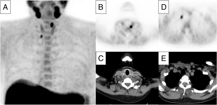
F-fluorocholine (FCH) PET/CT is a promising technique for visualizing hyperfunctioning parathyroid glands in hyperparathyroidism. It is still under debate whether to use this technique as a first-line imaging modality or to use it when conventional techniques such as Tc-sestamibi scintigraphy or ultrasonography are inconclusive. This study evaluates FCH PET/CT as a first-line modality.
Patients with primary hyperparathyroidism, referred between June 2015 and December 2018 for FCH PET/CT as a first-line imaging method, were included in this study. Baseline characteristics, clinical data, scan results, and type of treatment were recorded. The rate of correct detection was calculated on a per patient-based and a per lesion-based analysis. The reference standard comprised histopathological results, intraoperative response to parathyroidectomy, and clinical follow-up.
Two hundred and seventy-one patients were included, of which 139 patients underwent parathyroidectomy, 48 were treated with calcimimetics, and 84 patients received further follow-up without active treatment. In the surgically treated group, a single adenoma was suspected in 127 scans, double adenoma in three scans, and one scan showed evidence of three hyperfunctioning glands. In eight scans, no lesions were visualized. A total of 154 parathyroid glands were surgically removed. The rate of correct detection was calculated at 96% and 90%, on a per patient-based and per lesion-based analysis, respectively.
This retrospective study in a large cohort shows high detection rates of FCH PET/CT in primary hyperparathyroidism, which is in accordance to literature. The use of FCH PET/CT as a first-line imaging modality in preoperative planning of parathyroid surgery may therefore be a suitable choice.
F-氟胆碱(FCH)PET/CT是一种用于可视化甲状旁腺功能亢进症中功能亢进甲状旁腺的有前景的技术。对于是将该技术用作一线成像方式,还是在诸如锝- sestamibi闪烁扫描术或超声检查等传统技术结果不明确时使用,仍存在争议。本研究评估FCH PET/CT作为一线成像方式的情况。
纳入2015年6月至2018年12月期间因FCH PET/CT作为一线成像方法而转诊的原发性甲状旁腺功能亢进症患者。记录基线特征、临床数据、扫描结果和治疗类型。基于每位患者和基于每个病灶的分析计算正确检测率。参考标准包括组织病理学结果、甲状旁腺切除术中的反应以及临床随访。
共纳入271例患者,其中139例患者接受了甲状旁腺切除术,48例接受了拟钙剂治疗,84例患者接受进一步随访但未进行积极治疗。在手术治疗组中,127次扫描怀疑为单个腺瘤型,3次扫描怀疑为双腺瘤型,1次扫描显示有3个功能亢进腺体的证据。8次扫描未发现病变。共手术切除154个甲状旁腺。基于每位患者和基于每个病灶的分析计算的正确检测率分别为96%和90%。
这项对一大群患者的回顾性研究表明,FCH PET/CT在原发性甲状旁腺功能亢进症中的检测率很高,这与文献报道一致。因此在甲状旁腺手术的术前规划中,将FCH PET/CT用作一线成像方式可能是一个合适的选择。