• 文献检索
  • 文档翻译
  • 深度研究
  • 学术资讯
  • Suppr Zotero 插件Zotero 插件
  • 邀请有礼
  • 套餐&价格
  • 历史记录
应用&插件
Suppr Zotero 插件Zotero 插件浏览器插件Mac 客户端Windows 客户端微信小程序
定价
高级版会员购买积分包购买API积分包
服务
文献检索文档翻译深度研究API 文档MCP 服务
关于我们
关于 Suppr公司介绍联系我们用户协议隐私条款
关注我们

Suppr 超能文献

核心技术专利:CN118964589B侵权必究
粤ICP备2023148730 号-1Suppr @ 2026

文献检索

告别复杂PubMed语法,用中文像聊天一样搜索,搜遍4000万医学文献。AI智能推荐,让科研检索更轻松。

立即免费搜索

文件翻译

保留排版,准确专业,支持PDF/Word/PPT等文件格式,支持 12+语言互译。

免费翻译文档

深度研究

AI帮你快速写综述,25分钟生成高质量综述,智能提取关键信息,辅助科研写作。

立即免费体验

原发性甲状旁腺疾病诊断与管理的影像学建议:综述

Imaging Recommendations for Diagnosis and Management of Primary Parathyroid Pathologies: A Comprehensive Review.

作者信息

Chakrabarty Nivedita, Mahajan Abhishek, Basu Sandip, D'Cruz Anil K

机构信息

Department of Radiodiagnosis, Advanced Centre for Treatment, Research and Education in Cancer (ACTREC), Tata Memorial Centre, Homi Bhabha National Institute (HBNI), Parel, Mumbai 400012, Maharashtra, India.

Department of Imaging, The Clatterbridge Cancer Centre NHS Foundation Trust, 65 Pembroke Place, Liverpool L7 8YA, UK.

出版信息

Cancers (Basel). 2024 Jul 19;16(14):2593. doi: 10.3390/cancers16142593.

DOI:10.3390/cancers16142593
PMID:39061231
原文链接:https://pmc.ncbi.nlm.nih.gov/articles/PMC11274996/
Abstract

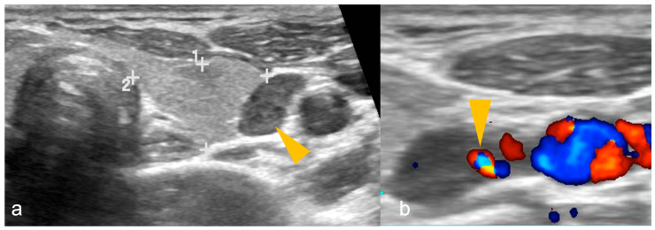
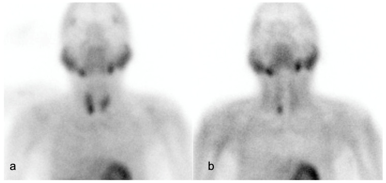
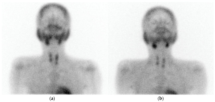
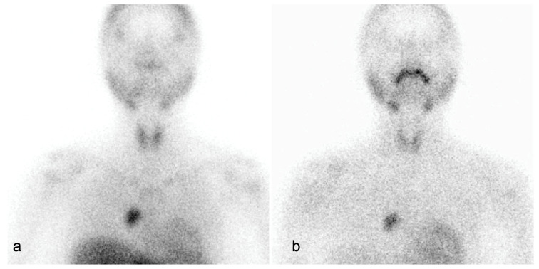
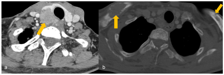
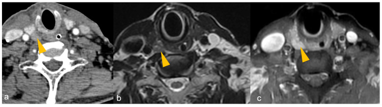
Parathyroid pathologies are suspected based on the biochemical alterations and clinical manifestations, and the predominant roles of imaging in primary hyperparathyroidism are localisation of tumour within parathyroid glands, surgical planning, and to look for any ectopic parathyroid tissue in the setting of recurrent disease. This article provides a comprehensive review of embryology and anatomical variations of parathyroid glands and their clinical relevance, surgical anatomy of parathyroid glands, differentiation between multiglandular parathyroid disease, solitary adenoma, atypical parathyroid tumour, and parathyroid carcinoma. The roles, advantages and limitations of ultrasound, four-dimensional computed tomography (4DCT), radiolabelled technetium-99 (Tc) sestamibi or dual tracer Tc pertechnetate and Tc-sestamibi with or without single photon emission computed tomography (SPECT) or SPECT/CT, dynamic enhanced magnetic resonance imaging (4DMRI), and fluoro-choline positron emission tomography (F-FCH PET) or [C] Methionine (C -MET) PET in the management of parathyroid lesions have been extensively discussed in this article. The role of fluorodeoxyglucose PET (FDG-PET) has also been elucidated in this article. Management guidelines for parathyroid carcinoma proposed by the American Society of Clinical Oncology (ASCO) have also been described. An algorithm for management of parathyroid lesions has been provided at the end to serve as a quick reference guide for radiologists, clinicians and surgeons.

摘要

基于生化改变和临床表现怀疑存在甲状旁腺病变,影像学在原发性甲状旁腺功能亢进中的主要作用是甲状旁腺肿瘤的定位、手术规划以及在复发性疾病情况下寻找任何异位甲状旁腺组织。本文全面综述了甲状旁腺的胚胎学、解剖变异及其临床相关性、甲状旁腺的手术解剖、多腺体甲状旁腺疾病、孤立性腺瘤、非典型甲状旁腺肿瘤和甲状旁腺癌之间的鉴别。本文还广泛讨论了超声、四维计算机断层扫描(4DCT)、放射性标记的锝-99(Tc)甲氧基异丁基异腈或双示踪剂高锝酸盐和Tc-甲氧基异丁基异腈联合或不联合单光子发射计算机断层扫描(SPECT)或SPECT/CT、动态增强磁共振成像(4DMRI)以及氟代胆碱正电子发射断层扫描(F-FCH PET)或[C]蛋氨酸(C-MET)PET在甲状旁腺病变管理中的作用、优势和局限性。本文还阐明了氟脱氧葡萄糖PET(FDG-PET)的作用。文中还描述了美国临床肿瘤学会(ASCO)提出的甲状旁腺癌管理指南。文末提供了甲状旁腺病变管理算法,作为放射科医生、临床医生和外科医生的快速参考指南。

https://cdn.ncbi.nlm.nih.gov/pmc/blobs/0431/11274996/fe88fcb559ad/cancers-16-02593-g007.jpg
https://cdn.ncbi.nlm.nih.gov/pmc/blobs/0431/11274996/8894b5dd9440/cancers-16-02593-g001.jpg
https://cdn.ncbi.nlm.nih.gov/pmc/blobs/0431/11274996/4b350dddb868/cancers-16-02593-g002.jpg
https://cdn.ncbi.nlm.nih.gov/pmc/blobs/0431/11274996/df8f13ddc07b/cancers-16-02593-g003.jpg
https://cdn.ncbi.nlm.nih.gov/pmc/blobs/0431/11274996/0b493d5cb1c5/cancers-16-02593-g004.jpg
https://cdn.ncbi.nlm.nih.gov/pmc/blobs/0431/11274996/e2eeb7a2afc3/cancers-16-02593-g005.jpg
https://cdn.ncbi.nlm.nih.gov/pmc/blobs/0431/11274996/f20070dd3d80/cancers-16-02593-g006.jpg
https://cdn.ncbi.nlm.nih.gov/pmc/blobs/0431/11274996/fe88fcb559ad/cancers-16-02593-g007.jpg

相似文献

1
Imaging Recommendations for Diagnosis and Management of Primary Parathyroid Pathologies: A Comprehensive Review.原发性甲状旁腺疾病诊断与管理的影像学建议:综述
Cancers (Basel). 2024 Jul 19;16(14):2593. doi: 10.3390/cancers16142593.
2
A Pilot Comparison of 18F-fluorocholine PET/CT, Ultrasonography and 123I/99mTc-sestaMIBI Dual-Phase Dual-Isotope Scintigraphy in the Preoperative Localization of Hyperfunctioning Parathyroid Glands in Primary or Secondary Hyperparathyroidism: Influence of Thyroid Anomalies.18F-氟胆碱PET/CT、超声检查及123I/99mTc-甲氧基异丁基异腈双相双同位素闪烁扫描术在原发性或继发性甲状旁腺功能亢进症中功能亢进甲状旁腺术前定位的初步比较:甲状腺异常的影响
Medicine (Baltimore). 2015 Oct;94(41):e1701. doi: 10.1097/MD.0000000000001701.
3
F-Fluorocholine PET/CT Is More Sensitive Than C-Methionine PET/CT for the Localization of Hyperfunctioning Parathyroid Tissue in Primary Hyperparathyroidism.在原发性甲状旁腺功能亢进症中,F-氟胆碱PET/CT在定位功能亢进的甲状旁腺组织方面比C-蛋氨酸PET/CT更敏感。
J Nucl Med. 2022 May;63(5):785-791. doi: 10.2967/jnumed.121.262395. Epub 2021 Aug 19.
4
F-Fluorocholine PET/CT in the assessment of primary hyperparathyroidism compared with Tc-MIBI or Tc-tetrofosmin SPECT/CT: a prospective dual-centre study in 100 patients.氟代胆碱 PET/CT 与 Tc-MIBI 或 Tc-四氮茂 SPECT/CT 评估原发性甲状旁腺功能亢进症的比较:100 例患者的前瞻性双中心研究。
Eur J Nucl Med Mol Imaging. 2018 Sep;45(10):1762-1771. doi: 10.1007/s00259-018-3980-9. Epub 2018 Mar 8.
5
Preoperative F-18 fluorocholine PET/CT for the detection of hyperfunctioning parathyroid glands in patients with secondary or tertiary hyperparathyroidism: comparison with Tc-99m sestamibi scan and neck ultrasound.术前 F-18 氟胆碱 PET/CT 检测继发性或三发性甲状旁腺功能亢进症患者的高功能甲状旁腺:与 Tc-99m sestamibi 扫描和颈部超声的比较。
Ann Nucl Med. 2020 Aug;34(8):527-537. doi: 10.1007/s12149-020-01479-2. Epub 2020 May 20.
6
Comparison of Neck Ultrasonography, Dual Phase Tc-Sestamibi with early SPECT-CT & F-Fluorocholine PET-CT as First Line Imaging in Patients with Primary Hyperparathyroidism.颈部超声、双相锝- sestamibi联合早期SPECT-CT与F-氟胆碱PET-CT作为原发性甲状旁腺功能亢进患者一线成像检查的比较
Indian J Nucl Med. 2023 Jul-Sep;38(3):208-217. doi: 10.4103/ijnm.ijnm_28_22. Epub 2023 Oct 10.
7
Is 18F-fluorocholine-positron emission tomography/computerized tomography a new imaging tool for detecting hyperfunctioning parathyroid glands in primary or secondary hyperparathyroidism?18F-氟胆碱正电子发射断层扫描/计算机断层扫描是否为原发性或继发性甲状旁腺功能亢进症中检测功能亢进甲状旁腺的新成像工具?
J Clin Endocrinol Metab. 2014 Dec;99(12):4531-6. doi: 10.1210/jc.2014-2821.
8
PETC/CT with F-Choline localizes hyperfunctioning parathyroid adenomas equally well in normocalcemic hyperparathyroidism as in overt hyperparathyroidism.F-胆碱 PETC/CT 同样能准确定位正常血钙型甲状旁腺功能亢进症和显性甲状旁腺功能亢进症中功能亢进的甲状旁腺腺瘤。
J Endocrinol Invest. 2019 Apr;42(4):419-426. doi: 10.1007/s40618-018-0931-z. Epub 2018 Aug 9.
9
18F-fluorocholine PET/MRI versus ultrasound and sestamibi for the localization of parathyroid adenomas.18F-氟胆碱 PET/MRI 与超声和 sestamibi 对甲状旁腺腺瘤定位的比较。
Langenbecks Arch Surg. 2023 Apr 20;408(1):155. doi: 10.1007/s00423-023-02893-6.
10
Added value of 18F-fluorocholine positron emission tomography-computed tomography in presurgical localization of hyperfunctioning parathyroid glands after dual tracer subtraction scintigraphy failure: A retrospective study of 47 patients.双示踪剂减影闪烁扫描失败后,18F-氟胆碱正电子发射断层扫描-计算机断层扫描在功能亢进甲状旁腺术前定位中的附加价值:47例患者的回顾性研究
Medicine (Baltimore). 2020 Jan;99(2):e18681. doi: 10.1097/MD.0000000000018681.

引用本文的文献

1
Is the F-choline PET-CT more cost-effective than standard protocol for locating a parathyroid adenoma?F-胆碱PET-CT在定位甲状旁腺腺瘤方面是否比标准方案更具成本效益?
Langenbecks Arch Surg. 2025 Jul 31;410(1):235. doi: 10.1007/s00423-025-03824-3.
2
[Parathyroid glands and their anatomical positional variations : Preoperative diagnostics and surgical planning].[甲状旁腺及其解剖位置变异:术前诊断与手术规划]
Chirurgie (Heidelb). 2025 Jul 24. doi: 10.1007/s00104-025-02343-7.
3
Human Chorionic Gonadotropin as a Marker of Parathyroid Carcinoma: Findings From a Scoping Review.

本文引用的文献

1
Normal Parathyroid Glands Are Most Often Seen on Neck Ultrasound.正常甲状旁腺在颈部超声中最常被看到。
Acad Radiol. 2024 Sep;31(9):3605-3611. doi: 10.1016/j.acra.2024.03.026. Epub 2024 May 22.
2
Four-dimensional computed tomography as first-line imaging in primary hyperparathyroidism, a retrospective comparison to conventional imaging in a predominantly single adenoma population.四维计算机断层扫描作为原发性甲状旁腺功能亢进的一线成像:在以单一腺瘤为主的人群中与传统成像的回顾性比较
EJNMMI Rep. 2024 May 1;8(1):11. doi: 10.1186/s41824-024-00198-5.
3
Imaging Analytics using Artificial Intelligence in Oncology: A Comprehensive Review.
人绒毛膜促性腺激素作为甲状旁腺癌的标志物:一项范围综述的结果
Cureus. 2025 Jun 16;17(6):e86141. doi: 10.7759/cureus.86141. eCollection 2025 Jun.
4
Normocalcemic Presentation of a Giant Polycystic Parathyroid Carcinoma.巨大多囊性甲状旁腺癌的血钙正常表现
JCEM Case Rep. 2025 Apr 18;3(6):luaf077. doi: 10.1210/jcemcr/luaf077. eCollection 2025 Jun.
5
Radiologist- and Surgeon-Performed Ultrasound (RSUS) Facilitates Minimally İnvasive Parathyroidectomy (MIP): Optimal Biochemical Parameters and Patient Outcomes.放射科医生和外科医生实施的超声检查(RSUS)有助于微创甲状旁腺切除术(MIP):最佳生化参数和患者预后。
J Clin Med. 2025 Mar 27;14(7):2279. doi: 10.3390/jcm14072279.
人工智能在肿瘤学中的影像分析:全面综述。
Clin Oncol (R Coll Radiol). 2024 Aug;36(8):498-513. doi: 10.1016/j.clon.2023.09.013. Epub 2023 Sep 27.
4
Can we skip technetium-99 m sestamibi scintigraphy in pediatric primary hyperparathyroidism patients with positive neck ultrasound results?对于颈部超声检查阳性的儿童原发性甲状旁腺功能亢进症患者,我们能否跳过锝-99m sestamibi 闪烁显像检查?
Pediatr Radiol. 2023 Oct;53(11):2253-2259. doi: 10.1007/s00247-023-05702-w. Epub 2023 Jul 13.
5
Selective venous sampling in primary hyperparathyroidism caused by ectopic parathyroid gland: a case report and literature review.原发性甲状旁腺功能亢进症中异位甲状旁腺引起的选择性静脉采血:病例报告及文献复习。
BMC Endocr Disord. 2023 Jul 6;23(1):141. doi: 10.1186/s12902-023-01376-5.
6
Preoperative imaging for hyperparathyroidism often takes upper parathyroid adenomas for lower adenomas.甲状旁腺功能亢进的术前影像学检查常将上甲状旁腺腺瘤误诊为下腺瘤。
Sci Rep. 2023 May 9;13(1):7568. doi: 10.1038/s41598-023-32707-0.
7
Normohormonal primary hyperparathyroidism: A systematic review and meta-analysis.正常激素原发性甲状旁腺功能亢进症:系统评价和荟萃分析。
Am J Surg. 2023 Aug;226(2):186-196. doi: 10.1016/j.amjsurg.2023.04.004. Epub 2023 Apr 19.
8
Multi-modality parathyroid imaging: A shifting paradigm.多模态甲状旁腺成像:一种不断变化的模式。
World J Radiol. 2023 Mar 28;15(3):69-82. doi: 10.4329/wjr.v15.i3.69.
9
Opportunistic CT Assessment of Parathyroid Glands: Utility of Radiologist-Recommended Biochemical Evaluation for Diagnosing Primary Hyperparathyroidism.甲状旁腺的机会性CT评估:放射科医生推荐的生化评估对诊断原发性甲状旁腺功能亢进的效用
AJR Am J Roentgenol. 2023 Aug;221(2):218-227. doi: 10.2214/AJR.23.29049. Epub 2023 Mar 22.
10
Comparison of 4D computed tomography and F-18 fluorocholine PET for localisation of parathyroid lesions in primary hyperparathyroidism: A systematic review and meta-analysis.4D 计算机断层扫描与 F-18 氟代胆碱 PET 对原发性甲状旁腺功能亢进症甲状旁腺病变定位的比较:系统评价和荟萃分析。
Clin Endocrinol (Oxf). 2023 Sep;99(3):262-271. doi: 10.1111/cen.14875. Epub 2023 Jan 8.