Department of Electronics and Information Engineering, Korea University, Sejong, Republic of Korea.
Department of Psychiatry, Korea University Guro Hospital, Korea University College of Medicine, Seoul, Republic of Korea.
PLoS One. 2019 Aug 1;14(8):e0220739. doi: 10.1371/journal.pone.0220739. eCollection 2019.
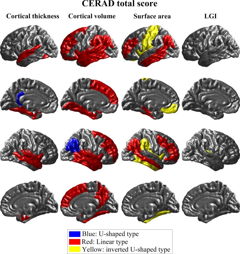
Several metrics of analysis of magnetic resonance imaging (MRI) have been used to assess Alzheimer's disease (AD)-related neurodegeneration. We compared four structural brain MRI analysis metrics, cortical thickness, volume, surface area, and local gyrification index (LGI), in different stages of AD-related cognitive decline. Participants with normal cognition, mild cognitive impairment, and AD were included (34 participants per group). All undertook the Consortium to Establish a Registry for Alzheimer's Disease (CERAD) battery of neuropsychological tests and brain MRI scanning. We analyzed associations between morphometric measures and CERAD total/ Mini Mental State Examination (MMSE) scores for the regions of interest (ROIs), identifying three types of curves: U-shaped, inverted U-shaped, and linear. Cortical thickness and volume analyses showed linear types in most of the significant ROIs. Significant ROIs for the cortical thickness analysis were located in the temporal and limbic lobes, whereas those for volume and surface area were distributed over more diffuse areas of the brain. LGI analysis showed few significant ROIs. CERAD total scores were more sensitive to early changes of cortical structures than MMSE scores. Cortical thickness analysis may be preferable in assessing brain structural MRI changes during AD-related cognitive decline, whereas LGI analysis may have limited capability to reflect the cognitive decrease. Our findings may provide a reference for future studies and help to establish optimal analytical approaches to brain structural MRI in neurodegenerative diseases.
几种磁共振成像(MRI)分析指标已被用于评估与阿尔茨海默病(AD)相关的神经退行性变。我们比较了四种结构性脑 MRI 分析指标,即皮质厚度、体积、表面积和局部脑回指数(LGI),以评估 AD 相关认知下降的不同阶段。参与者包括认知正常、轻度认知障碍和 AD 患者(每组 34 名参与者)。所有参与者都接受了 Consortium to Establish a Registry for Alzheimer's Disease(CERAD)神经心理学测试和大脑 MRI 扫描。我们分析了形态测量指标与 CERAD 总/简易精神状态检查(MMSE)评分之间的关联,确定了三种类型的曲线:U 型、倒 U 型和线性。皮质厚度和体积分析在大多数显著 ROI 中显示出线性类型。皮质厚度分析的显著 ROI 位于颞叶和边缘叶,而体积和表面积分析的 ROI 分布在大脑更弥散的区域。LGI 分析显示出较少的显著 ROI。CERAD 总评分比 MMSE 评分更能敏感地反映皮质结构的早期变化。皮质厚度分析可能更适合评估 AD 相关认知下降期间的大脑结构 MRI 变化,而 LGI 分析可能无法反映认知下降。我们的研究结果可能为未来的研究提供参考,并有助于建立神经退行性疾病中大脑结构 MRI 的最佳分析方法。