• 文献检索
  • 文档翻译
  • 深度研究
  • 学术资讯
  • Suppr Zotero 插件Zotero 插件
  • 邀请有礼
  • 套餐&价格
  • 历史记录
应用&插件
Suppr Zotero 插件Zotero 插件浏览器插件Mac 客户端Windows 客户端微信小程序
定价
高级版会员购买积分包购买API积分包
服务
文献检索文档翻译深度研究API 文档MCP 服务
关于我们
关于 Suppr公司介绍联系我们用户协议隐私条款
关注我们

Suppr 超能文献

核心技术专利:CN118964589B侵权必究
粤ICP备2023148730 号-1Suppr @ 2026

文献检索

告别复杂PubMed语法,用中文像聊天一样搜索,搜遍4000万医学文献。AI智能推荐,让科研检索更轻松。

立即免费搜索

文件翻译

保留排版,准确专业,支持PDF/Word/PPT等文件格式,支持 12+语言互译。

免费翻译文档

深度研究

AI帮你快速写综述,25分钟生成高质量综述,智能提取关键信息,辅助科研写作。

立即免费体验

从眶上小切口、额内侧和翼点入路视角看视交叉间、经终板、视颈动脉三角及颈动脉池窗:一项带人工损伤的尸体对照研究

Interoptic, Trans-lamina Terminalis, Opticocarotid Triangle, and Caroticosylvian Windows From Mini-Supraorbital, Frontomedial, and Pterional Perspectives: A Comparative Cadaver Study With Artificial Lesions.

作者信息

Adib Sasan Darius, Herlan Stephan, Ebner Florian H, Hirt Bernhard, Tatagiba Marcos, Honegger Juergen

机构信息

Department of Neurosurgery, University of Tübingen, Tübingen, Germany.

Department of Clinical Anatomy and Cell Analysis, University of Tübingen, Tübingen, Germany.

出版信息

Front Surg. 2019 Jul 16;6:40. doi: 10.3389/fsurg.2019.00040. eCollection 2019.

DOI:10.3389/fsurg.2019.00040
PMID:31380387
原文链接:https://pmc.ncbi.nlm.nih.gov/articles/PMC6646665/
Abstract

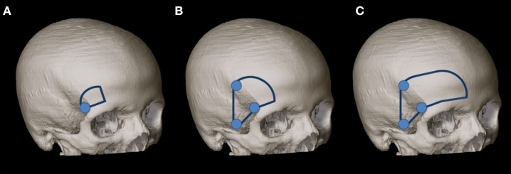
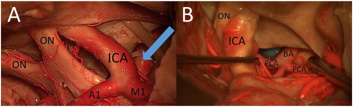
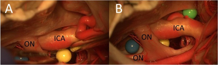
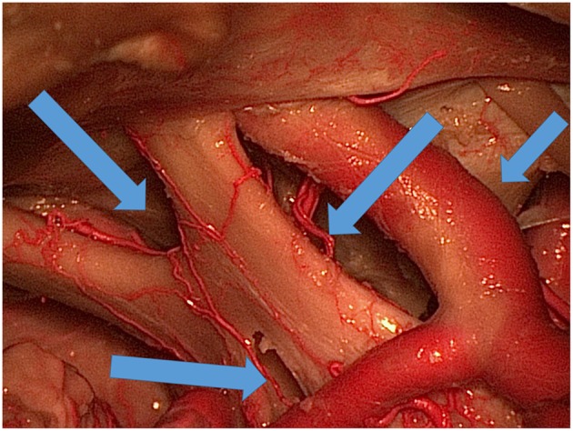
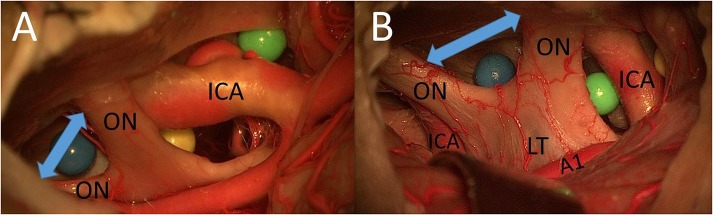
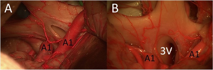
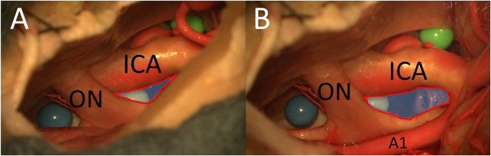
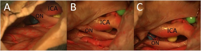
The mini-supraorbital (MSO) and pterional (PT) approaches have been compared in a number of studies focusing on the treatment of aneurysms, craniopharyngiomas, and meningiomas. The goal of this study was to analyze the surgical exposure to different artificial lesions through interoptic (IO), trans-lamina terminalis (TLT), opticocarotid triangle (OCT), and caroticosylvian (CS) windows from the MSO, frontomedial (FM), and PT perspectives. The MSO, PT, and FM approaches were performed sequentially in two fixed cadaver heads. Three colored spheres were placed around the optic chiasm: (1) between the optic nerves; (2) between the optic nerve and the internal carotid artery; and (3) between the internal carotid artery and the oculomotor nerve. The surgical exposures to these structures by using the IO, TLT, OCT, and CS windows were compared. (1) IO window: from the MSO and PT approaches, the total surgical exposure mainly allows visualization of contralateral lesions. The FM approach was superior for exploration of both sides of the area between the optic nerves. (2) TLT pathway: the MSO and PT approaches mainly expose the contralateral third ventricle wall. (3) OCT window: the PT approach allows exposure of a larger part of the sphere between the optic nerve and the internal carotid artery than the MSO approach. (4) CS window: the PT approach allows a better exposure of lateral structures such as the oculomotor nerve and of the medial prepontine area in comparison to the MSO approach. Simulation of the surgical situation with artificial lesions is a good model for comparing surgical perspectives and for analyzing feasibility of lesion exposure and resection.

摘要

在许多针对动脉瘤、颅咽管瘤和脑膜瘤治疗的研究中,已对眶上缘内侧(MSO)入路和翼点(PT)入路进行了比较。本研究的目的是从MSO、额内侧(FM)和PT视角,分析通过视交叉间(IO)、终板经路(TLT)、视颈三角(OCT)和颈内动脉-大脑外侧裂(CS)窗口对不同人工病灶的手术显露情况。在两个固定的尸体头部依次进行MSO、PT和FM入路。在视交叉周围放置三个彩色球体:(1)位于视神经之间;(2)位于视神经与颈内动脉之间;(3)位于颈内动脉与动眼神经之间。比较使用IO、TLT、OCT和CS窗口对这些结构的手术显露情况。(1)IO窗口:从MSO和PT入路来看,总的手术显露主要能看到对侧病灶。FM入路在探查视神经之间区域的两侧方面更具优势。(2)TLT路径:MSO和PT入路主要显露对侧第三脑室壁。(3)OCT窗口:与MSO入路相比,PT入路能显露视神经与颈内动脉之间球体的更大一部分。(4)CS窗口:与MSO入路相比,PT入路能更好地显露动眼神经等外侧结构以及脑桥前内侧区域。用人工病灶模拟手术情况是比较手术视角以及分析病灶显露和切除可行性的良好模型。

https://cdn.ncbi.nlm.nih.gov/pmc/blobs/eb8d/6646665/cca61ffb1297/fsurg-06-00040-g0008.jpg
https://cdn.ncbi.nlm.nih.gov/pmc/blobs/eb8d/6646665/92101385405f/fsurg-06-00040-g0001.jpg
https://cdn.ncbi.nlm.nih.gov/pmc/blobs/eb8d/6646665/24c985e9713f/fsurg-06-00040-g0002.jpg
https://cdn.ncbi.nlm.nih.gov/pmc/blobs/eb8d/6646665/557b86e7d4cf/fsurg-06-00040-g0003.jpg
https://cdn.ncbi.nlm.nih.gov/pmc/blobs/eb8d/6646665/3f6ea52aba4c/fsurg-06-00040-g0004.jpg
https://cdn.ncbi.nlm.nih.gov/pmc/blobs/eb8d/6646665/524e7a50837c/fsurg-06-00040-g0005.jpg
https://cdn.ncbi.nlm.nih.gov/pmc/blobs/eb8d/6646665/bf998765785d/fsurg-06-00040-g0006.jpg
https://cdn.ncbi.nlm.nih.gov/pmc/blobs/eb8d/6646665/384d93c31bf1/fsurg-06-00040-g0007.jpg
https://cdn.ncbi.nlm.nih.gov/pmc/blobs/eb8d/6646665/cca61ffb1297/fsurg-06-00040-g0008.jpg

相似文献

1
Interoptic, Trans-lamina Terminalis, Opticocarotid Triangle, and Caroticosylvian Windows From Mini-Supraorbital, Frontomedial, and Pterional Perspectives: A Comparative Cadaver Study With Artificial Lesions.从眶上小切口、额内侧和翼点入路视角看视交叉间、经终板、视颈动脉三角及颈动脉池窗:一项带人工损伤的尸体对照研究
Front Surg. 2019 Jul 16;6:40. doi: 10.3389/fsurg.2019.00040. eCollection 2019.
2
Pre- versus post-anterior clinoidectomy measurements of the optic nerve, internal carotid artery, and opticocarotid triangle: a cadaveric morphometric study.前床突切除术前后视神经、颈内动脉及视神经-颈动脉三角的测量:一项尸体形态学研究
Neurosurgery. 2000 Apr;46(4):1018-21; discussion 1021-3.
3
Surgical limits in transnasal approach to opticocarotid region and planum sphenoidale: an anatomic cadaveric study.经鼻入路视神经-颈内动脉区和蝶骨平台手术界限:尸体解剖研究
World Neurosurg. 2010 Apr;73(4):326-33. doi: 10.1016/j.wneu.2010.01.015.
4
From above or from below? That is the question. Comparison of the supraorbital approach with the endonasal approach. A cadaveric study.从上方还是从下方?这就是问题所在。眶上入路与鼻内入路的比较。一项尸体研究。
Br J Neurosurg. 2018 Oct;32(5):548-552. doi: 10.1080/02688697.2018.1480748. Epub 2018 Jun 6.
5
Quantitative anatomical analysis and clinical experience with mini-pterional and mini-orbitozygomatic approaches for intracranial aneurysm surgery.经翼点和眶颧微型入路治疗颅内动脉瘤的定量解剖分析及临床经验。
J Neurosurg. 2017 Sep;127(3):646-659. doi: 10.3171/2016.6.JNS16306. Epub 2016 Nov 18.
6
Lateral supraorbital approach vs pterional approach: an anatomic qualitative and quantitative evaluation.外侧眶上入路与翼点入路:解剖学定性和定量评估。
Neurosurgery. 2011 Jun;68(2 Suppl Operative):364-72; discussion 371-2. doi: 10.1227/NEU.0b013e318211721f.
7
The approach angle to the interoptic triangle limits surgical workspace when targeting the contralateral internal carotid artery.视交叉三角入路限制了对侧颈内动脉手术空间。
Acta Neurochir (Wien). 2019 Aug;161(8):1535-1543. doi: 10.1007/s00701-019-03911-7. Epub 2019 May 18.
8
Quantitative study of the opticocarotid and carotid-oculomotor windows for the interpeduncular fossa, before and after internal carotid artery mobilization and posterior communicating division.颈内动脉游离和后交通动脉分支前后,视交叉颈动脉窗和颈动脉动眼神经窗对脚间窝的定量研究。
Neurosurgery. 2015 Mar;11 Suppl 2:162-79; discussion 179-80. doi: 10.1227/NEU.0000000000000617.
9
The endoscopic supraorbital translaminar approach: a technical note.内镜眶上锁孔入路:技术说明。
Acta Neurochir (Wien). 2021 Mar;163(3):635-641. doi: 10.1007/s00701-020-04498-0. Epub 2020 Jul 20.
10
Using the trans-lamina terminalis route via a pterional approach to resect a retrochiasmatic craniopharyngioma involving the third ventricle.采用经翼点入路经终板间隙切除累及第三脑室的视交叉后颅咽管瘤。
Neurosurg Focus. 2016 Jan;40 Video Suppl 1:2016.1.FocusVid.15440. doi: 10.3171/2016.1.FocusVid.15440.

引用本文的文献

1
Petroclival Clinoidal Folds and Arachnoidal Membranes of the Anteromedial Incisural Space: Clinical Anatomy for Neuro Critical Care.前内侧脑裂间隙的岩斜床突皱襞和蛛网膜:神经重症监护的临床解剖学
Diagnostics (Basel). 2023 Oct 13;13(20):3203. doi: 10.3390/diagnostics13203203.
2
Microneurosurgical anatomy of the basal cisterns: A brief review for cisternostomy.脑池的显微神经外科解剖:脑脊液分流术的简要综述
Surg Neurol Int. 2023 Mar 24;14:97. doi: 10.25259/SNI_1095_2022. eCollection 2023.

本文引用的文献

1
Microsurgical resection of tumors of the lateral and third ventricles: operative corridors for difficult-to-reach lesions.侧脑室和第三脑室肿瘤的显微手术切除:针对难以触及病变的手术入路
J Neurooncol. 2016 Nov;130(2):331-340. doi: 10.1007/s11060-016-2126-9. Epub 2016 May 27.
2
Optic nerve mobilization to enhance the exposure of the pituitary stalk during craniopharyngioma resection: early experience.视神经牵拉术增强颅咽管瘤切除术中垂体柄显露:初步经验。
J Neurosurg. 2016 Sep;125(3):683-8. doi: 10.3171/2015.6.JNS141847. Epub 2015 Dec 18.
3
Contralateral Approach to Internal Carotid Artery Ophthalmic Segment Aneurysms: Angiographic Analysis and Surgical Results for 30 Patients.
颈内动脉眼段动脉瘤的对侧入路:30例患者的血管造影分析及手术结果
Neurosurgery. 2015 Jul;77(1):104-12; discussion 112. doi: 10.1227/NEU.0000000000000742.
4
Quantitative study of the opticocarotid and carotid-oculomotor windows for the interpeduncular fossa, before and after internal carotid artery mobilization and posterior communicating division.颈内动脉游离和后交通动脉分支前后,视交叉颈动脉窗和颈动脉动眼神经窗对脚间窝的定量研究。
Neurosurgery. 2015 Mar;11 Suppl 2:162-79; discussion 179-80. doi: 10.1227/NEU.0000000000000617.
5
Intracranial injectable tumor model: technical advancements.颅内注射肿瘤模型:技术进展
J Neurol Surg B Skull Base. 2014 Oct;75(5):301-8. doi: 10.1055/s-0034-1368148. Epub 2014 Jul 21.
6
Microsurgical resection of extensive craniopharyngiomas using a frontolateral approach: operative technique and outcome.经额眶颧入路显微切除术治疗巨大颅咽管瘤:手术技术与疗效。
J Neurosurg. 2014 Feb;120(2):559-70. doi: 10.3171/2013.9.JNS122133. Epub 2013 Nov 22.
7
Comparative Analysis of the Mini-pterional and Supraorbital Keyhole Craniotomies for Unruptured Aneurysms with Numeric Measurements of Their Geometric Configurations.经翼点微骨窗与眶上锁孔开颅术治疗未破裂动脉瘤的比较分析及其几何构型的数值测量
J Cerebrovasc Endovasc Neurosurg. 2013 Mar;15(1):5-12. doi: 10.7461/jcen.2013.15.1.5. Epub 2013 Mar 31.
8
Surgical treatment of ruptured anterior circulation aneurysms: comparison of pterional and supraorbital keyhole approaches.前循环破裂动脉瘤的手术治疗:翼点入路与眶上锁孔入路的比较。
Neurosurgery. 2013 Mar;72(3):437-41; discussion 441-2. doi: 10.1227/NEU.0b013e3182804e9c.
9
Quantitative verification of the keyhole concept: a comparison of area of exposure in the parasellar region via supraorbital keyhole, frontotemporal pterional, and supraorbital approaches.锁孔概念的定量验证:经眶上锁孔、额颞翼点和眶上锁孔入路蝶鞍旁区显露面积的比较。
J Neurosurg. 2013 Feb;118(2):264-9. doi: 10.3171/2012.9.JNS09186. Epub 2012 Nov 9.
10
Unruptured Supraclinoid Internal Carotid Artery Aneurysm Surgery : Superciliary Keyhole Approach versus Pterional Approach.未破裂的鞍上颈内动脉动脉瘤手术:眉弓锁孔入路与翼点入路对比
J Korean Neurosurg Soc. 2012 Oct;52(4):306-11. doi: 10.3340/jkns.2012.52.4.306. Epub 2012 Oct 22.