• 文献检索
  • 文档翻译
  • 深度研究
  • 学术资讯
  • Suppr Zotero 插件Zotero 插件
  • 邀请有礼
  • 套餐&价格
  • 历史记录
应用&插件
Suppr Zotero 插件Zotero 插件浏览器插件Mac 客户端Windows 客户端微信小程序
定价
高级版会员购买积分包购买API积分包
服务
文献检索文档翻译深度研究API 文档MCP 服务
关于我们
关于 Suppr公司介绍联系我们用户协议隐私条款
关注我们

Suppr 超能文献

核心技术专利:CN118964589B侵权必究
粤ICP备2023148730 号-1Suppr @ 2026

文献检索

告别复杂PubMed语法,用中文像聊天一样搜索,搜遍4000万医学文献。AI智能推荐,让科研检索更轻松。

立即免费搜索

文件翻译

保留排版,准确专业,支持PDF/Word/PPT等文件格式,支持 12+语言互译。

免费翻译文档

深度研究

AI帮你快速写综述,25分钟生成高质量综述,智能提取关键信息,辅助科研写作。

立即免费体验

心脏磁共振成像揭示的心外膜脂肪组织的侵入性本质

The Intrusive Nature of Epicardial Adipose Tissue as Revealed by Cardiac Magnetic Resonance.

作者信息

Leo Laura Anna, Paiocchi Vera Lucia, Schlossbauer Susanne Anna, Ho Siew Yen, Faletra Francesco F

机构信息

Cardiac Imaging Department, Fondazione Cardiocentro Ticino, Lugano, Switzerland.

Department of Cardiac Morphology, Royal Brompton Hospital and Imperial College London, London, UK.

出版信息

J Cardiovasc Echogr. 2019 Apr-Jun;29(2):45-51. doi: 10.4103/jcecho.jcecho_22_19.

DOI:10.4103/jcecho.jcecho_22_19
PMID:31392118
原文链接:https://pmc.ncbi.nlm.nih.gov/articles/PMC6657468/
Abstract

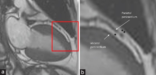
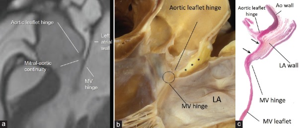
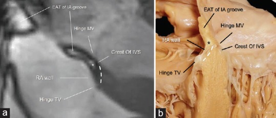
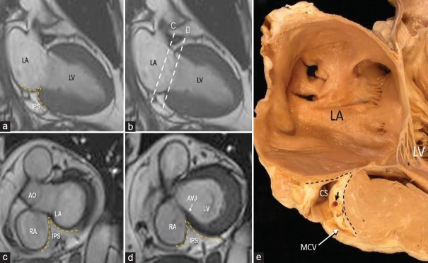
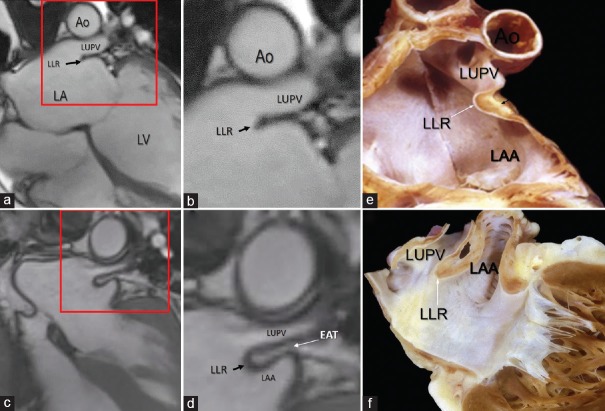
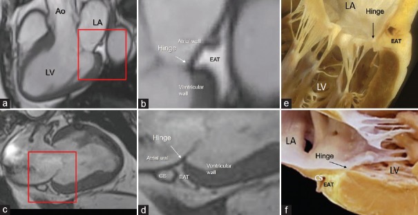
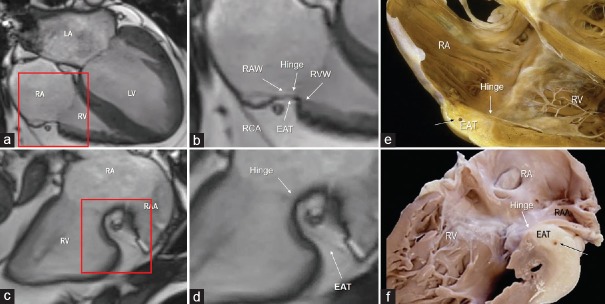
The epicardial adipose tissue (EAT) refers to the deposition of adipose tissue fully enclosed by the pericardial sac. EAT has a complex mixture of adipocytes, nervous tissue, as well as inflammatory, stromal and immune cells secreting bioactive molecules. This heterogeneous composition reveals that it is not a simply fat storage depot, but rather a biologically active organ that appears playing a "dichotomous" role, either protective or proinflammatory and proatherogenic. The cardiac magnetic resonance (CMR) allows a clear visualization of EAT using a specific pulse sequence called steady-state free precession. When abundant, the EAT assumes a pervasive presence not only covering the entire epicardial surface but also invading spaces that usually are almost virtual and separating walls that usually are so close each other to resemble a single wall. To the best of our knowledge, this aspect of cardiac anatomy has never been described before. In this pictorial review, we therefore focus our attention on certain cardiac areas in which EAT, when abundant, is particularly intrusive. In particular, we describe the presence of EAT into: (a) the interatrial groove, the atrioventricular septum, and the inferior pyramidal space, (b) the left lateral ridge, (c) the atrioventricular grooves, and (d) the transverse pericardial sinus. To confirm the reliability in depicting the EAT distribution, we present CMR images side-by-side with corresponding anatomic specimens.

摘要

心外膜脂肪组织(EAT)是指被心包囊完全包裹的脂肪组织沉积。EAT由脂肪细胞、神经组织以及分泌生物活性分子的炎症细胞、基质细胞和免疫细胞混合而成。这种异质性组成表明它并非简单的脂肪储存库,而是一个具有生物活性的器官,似乎发挥着“双重”作用,既有保护作用,也有促炎和促动脉粥样硬化作用。心脏磁共振成像(CMR)使用一种称为稳态自由进动的特定脉冲序列能够清晰显示EAT。当EAT大量存在时,它不仅会遍布整个心外膜表面,还会侵入通常几乎是虚拟的空间,并分隔通常彼此靠近而看似单一壁的壁。据我们所知,心脏解剖学的这一方面以前从未被描述过。因此,在这篇图片综述中,我们将注意力集中在某些心脏区域,当EAT大量存在时,这些区域的EAT特别具有侵入性。具体来说,我们描述了EAT在以下部位的存在情况:(a)房间沟、房室间隔和下锥体间隙,(b)左侧嵴,(c)房室沟,以及(d)心包横窦。为了证实描绘EAT分布的可靠性,我们将CMR图像与相应的解剖标本并排展示。

https://cdn.ncbi.nlm.nih.gov/pmc/blobs/67ea/6657468/4a0cf9916ea3/JCE-29-45-g008.jpg
https://cdn.ncbi.nlm.nih.gov/pmc/blobs/67ea/6657468/714fd9ee48d1/JCE-29-45-g001.jpg
https://cdn.ncbi.nlm.nih.gov/pmc/blobs/67ea/6657468/10c7f1eeff00/JCE-29-45-g002.jpg
https://cdn.ncbi.nlm.nih.gov/pmc/blobs/67ea/6657468/decc057a7e4f/JCE-29-45-g003.jpg
https://cdn.ncbi.nlm.nih.gov/pmc/blobs/67ea/6657468/4c5eec032f79/JCE-29-45-g004.jpg
https://cdn.ncbi.nlm.nih.gov/pmc/blobs/67ea/6657468/52367ec9be59/JCE-29-45-g005.jpg
https://cdn.ncbi.nlm.nih.gov/pmc/blobs/67ea/6657468/856df3cf4c59/JCE-29-45-g006.jpg
https://cdn.ncbi.nlm.nih.gov/pmc/blobs/67ea/6657468/9435a5d32a27/JCE-29-45-g007.jpg
https://cdn.ncbi.nlm.nih.gov/pmc/blobs/67ea/6657468/4a0cf9916ea3/JCE-29-45-g008.jpg

相似文献

1
The Intrusive Nature of Epicardial Adipose Tissue as Revealed by Cardiac Magnetic Resonance.心脏磁共振成像揭示的心外膜脂肪组织的侵入性本质
J Cardiovasc Echogr. 2019 Apr-Jun;29(2):45-51. doi: 10.4103/jcecho.jcecho_22_19.
2
Electroanatomical Remodeling of the Atria in Obesity: Impact of Adjacent Epicardial Fat.肥胖患者心房的电重构:心外膜脂肪的影响。
JACC Clin Electrophysiol. 2018 Dec;4(12):1529-1540. doi: 10.1016/j.jacep.2018.08.014. Epub 2018 Nov 1.
3
Bioimpedance analysis parameters and epicardial adipose tissue assessed by cardiac magnetic resonance imaging in patients with heart failure.心脏磁共振成像评估心力衰竭患者的生物阻抗分析参数和心外膜脂肪组织。
Obesity (Silver Spring). 2010 Dec;18(12):2326-32. doi: 10.1038/oby.2010.65. Epub 2010 Mar 25.
4
Role of Epicardial Adipose Tissue in Health and Disease: A Matter of Fat?心外膜脂肪组织在健康与疾病中的作用:脂肪问题?
Compr Physiol. 2017 Jun 18;7(3):1051-1082. doi: 10.1002/cphy.c160034.
5
The epicardial adipose tissue confined in the atrioventricular groove can be used to assess atrial adipose tissue and atrial dysfunction in cardiac magnetic resonance imaging.位于房室沟内的心外膜脂肪组织可用于在心脏磁共振成像中评估心房脂肪组织和心房功能障碍。
Eur Heart J Imaging Methods Pract. 2024 Jun 14;2(1):qyae057. doi: 10.1093/ehjimp/qyae057. eCollection 2024 Jan.
6
The relationship between location-specific epicardial adipose tissue volume and coronary atherosclerotic plaque burden in type 2 diabetic patients.2型糖尿病患者特定部位的心外膜脂肪组织体积与冠状动脉粥样硬化斑块负荷之间的关系。
Kardiol Pol. 2017;75(3):204-212. doi: 10.5603/KP.a2016.0167. Epub 2016 Dec 13.
7
Relations of epicardial adipose tissue measured by multidetector computed tomography to components of the metabolic syndrome are region-specific and independent of anthropometric indexes and intraabdominal visceral fat.通过多排螺旋计算机断层扫描测量的心外膜脂肪组织与代谢综合征各组分的关系具有区域特异性,且独立于人体测量指标和腹内内脏脂肪。
J Clin Endocrinol Metab. 2009 Feb;94(2):662-9. doi: 10.1210/jc.2008-0834. Epub 2008 Dec 2.
8
Epicardial adipose tissue: far more than a fat depot.心外膜脂肪组织:远不止是一个脂肪库。
Cardiovasc Diagn Ther. 2014 Dec;4(6):416-29. doi: 10.3978/j.issn.2223-3652.2014.11.05.
9
Evolutional change in epicardial fat and its correlation with myocardial diffuse fibrosis in heart failure patients.心力衰竭患者心外膜脂肪的演变及其与心肌弥漫性纤维化的相关性。
J Clin Lipidol. 2017 Nov-Dec;11(6):1421-1431. doi: 10.1016/j.jacl.2017.08.018. Epub 2017 Sep 4.
10
[Epicardial adipose tissue and its role in cardiac physiology and disease].[心外膜脂肪组织及其在心脏生理学和疾病中的作用]
Postepy Hig Med Dosw (Online). 2013 Jun 20;67:584-93. doi: 10.5604/17322693.1053908.

引用本文的文献

1
The Role of Imaging in Cardiovascular Prevention: A Comprehensive Review.影像学在心血管疾病预防中的作用:全面综述
J Cardiovasc Echogr. 2025 Jan-Mar;35(1):8-18. doi: 10.4103/jcecho.jcecho_26_25. Epub 2025 Apr 30.
2
Assessment of epicardial adipose tissue volume and carotid intima-media thickness in children with primary arterial hypertension by magnetic resonance imaging.通过磁共振成像评估原发性动脉高血压患儿的心外膜脂肪组织体积和颈动脉内膜中层厚度。
Radiol Oncol. 2025 May 14;59(2):1-10. doi: 10.2478/raon-2025-0030. eCollection 2025 Jun 1.
3
Cardiac magnetic resonance quantified epicardial fat volume is associated with complex coronary artery disease among diabetics.

本文引用的文献

1
Imaging-based tricuspid valve anatomy by computed tomography, magnetic resonance imaging, two and three-dimensional echocardiography: correlation with anatomic specimen.基于计算机断层扫描、磁共振成像、二维和三维超声心动图的三尖瓣成像:与解剖标本的相关性。
Eur Heart J Cardiovasc Imaging. 2019 Jan 1;20(1):1-13. doi: 10.1093/ehjci/jey136.
2
The role of epicardial adipose tissue in cardiac biology: classic concepts and emerging roles.心外膜脂肪组织在心脏生物学中的作用:经典概念与新出现的作用
J Physiol. 2017 Jun 15;595(12):3907-3917. doi: 10.1113/JP273049. Epub 2017 Mar 13.
3
Lipomatous Atrial Septal Hypertrophy: A Review of Its Anatomy, Pathophysiology, Multimodality Imaging, and Relevance to Percutaneous Interventions.
心脏磁共振定量的心外膜脂肪体积与糖尿病患者的复杂冠状动脉疾病相关。
Cardiovasc Diabetol. 2025 Feb 7;24(1):64. doi: 10.1186/s12933-025-02606-x.
4
Charting the Unseen: How Non-Invasive Imaging Could Redefine Cardiovascular Prevention.绘制不可见之物:无创成像如何重新定义心血管疾病预防
J Cardiovasc Dev Dis. 2024 Aug 9;11(8):245. doi: 10.3390/jcdd11080245.
5
Association between Epicardial Adipose Tissue and Atrial Fibrillation in Patients with Transfusion-Dependent β-Thalassemia.依赖输血的β地中海贫血患者的心外膜脂肪组织与心房颤动之间的关联
J Clin Med. 2024 Jun 14;13(12):3471. doi: 10.3390/jcm13123471.
6
The Role of Epicardial Adipose Tissue in Acute Coronary Syndromes, Post-Infarct Remodeling and Cardiac Regeneration.心外膜脂肪组织在急性冠状动脉综合征、心肌梗死后重构和心脏再生中的作用。
Int J Mol Sci. 2024 Mar 22;25(7):3583. doi: 10.3390/ijms25073583.
7
Role of epicardial adipose tissue in diabetic cardiomyopathy through the lens of cardiovascular magnetic resonance imaging - a narrative review.从心血管磁共振成像角度看心外膜脂肪组织在糖尿病性心肌病中的作用——一篇叙述性综述
Ther Adv Endocrinol Metab. 2024 Mar 10;15:20420188241229540. doi: 10.1177/20420188241229540. eCollection 2024.
8
Epicardial Adipose Tissue: A Piece of The Puzzle in Pediatric Hypertension.心外膜脂肪组织:小儿高血压难题中的一块拼图。
J Clin Med. 2023 Mar 12;12(6):2192. doi: 10.3390/jcm12062192.
9
Correlation analysis of epicardial adipose tissue volume quantified by computed tomography images and coronary heart disease under optimized reconstruction algorithm.基于优化重建算法的计算机断层扫描图像定量分析的心外膜脂肪组织体积与冠心病的相关性分析
Pak J Med Sci. 2021;37(6):1677-1681. doi: 10.12669/pjms.37.6-WIT.4882.
10
Subclinical Left Ventricular Dysfunction in Severe Obesity and Reverse Cardiac Remodeling after Bariatric Surgery.严重肥胖患者的亚临床左心室功能障碍及减重手术后的心脏逆向重塑
J Cardiovasc Echogr. 2020 Jan-Mar;30(1):22-28. doi: 10.4103/jcecho.jcecho_50_19. Epub 2020 Apr 13.
脂肪性房间隔肥厚:解剖学、病理生理学、多模态成像及其与经皮介入相关性的综述
J Am Soc Echocardiogr. 2016 Aug;29(8):717-723. doi: 10.1016/j.echo.2016.04.014. Epub 2016 Jun 7.
4
Interscan reproducibility of computer-aided epicardial and thoracic fat measurement from noncontrast cardiac CT.非增强心脏 CT 计算机辅助心外膜和胸腔脂肪测量的 Interscan 可重复性。
J Cardiovasc Comput Tomogr. 2011 May-Jun;5(3):172-9. doi: 10.1016/j.jcct.2011.03.009. Epub 2011 Mar 21.
5
The architecture of the left lateral atrial wall: a particular anatomic region with implications for ablation of atrial fibrillation.左心房外侧壁的结构:一个对房颤消融有影响的特殊解剖区域。
Eur Heart J. 2008 Feb;29(3):356-62. doi: 10.1093/eurheartj/ehm606.
6
Epicardial fat: properties, function and relationship to obesity.心外膜脂肪:特性、功能及其与肥胖的关系。
Obes Rev. 2007 May;8(3):253-61. doi: 10.1111/j.1467-789X.2006.00293.x.
7
Epicardial adipose tissue: anatomic, biomolecular and clinical relationships with the heart.心外膜脂肪组织:与心脏的解剖学、生物分子学及临床关系
Nat Clin Pract Cardiovasc Med. 2005 Oct;2(10):536-43. doi: 10.1038/ncpcardio0319.
8
The ventricular epicardial fat is related to the myocardial mass in normal, ischemic and hypertrophic hearts.在正常、缺血和肥厚型心脏中,心室心外膜脂肪与心肌质量相关。
Cardiovasc Pathol. 2004 Nov-Dec;13(6):313-6. doi: 10.1016/j.carpath.2004.08.005.
9
Human epicardial adipose tissue is a source of inflammatory mediators.人的心外膜脂肪组织是炎症介质的一个来源。
Circulation. 2003 Nov 18;108(20):2460-6. doi: 10.1161/01.CIR.0000099542.57313.C5. Epub 2003 Oct 27.
10
Epicardial fat from echocardiography: a new method for visceral adipose tissue prediction.超声心动图测量的心外膜脂肪:一种预测内脏脂肪组织的新方法。
Obes Res. 2003 Feb;11(2):304-10. doi: 10.1038/oby.2003.45.