Carerj Maria Ludovica, Restelli Davide, Poleggi Cristina, Di Bella Gianluca, Zito Concetta, Manganaro Roberta, Piccione Maurizio Cusmà, Trimarchi Giancarlo, Farina Andrea, Micari Antonio, Carerj Scipione
Department of Perioperative Cardiology and Cardiovascular Imaging, Centro Cardiologico Monzino IRCCS, Milan, Italy.
Department of Cardio-Thoraco-Vascular Care, ASST Lecco - Ospedale A. Manzoni, Lecco, Italy.
J Cardiovasc Echogr. 2025 Jan-Mar;35(1):8-18. doi: 10.4103/jcecho.jcecho_26_25. Epub 2025 Apr 30.
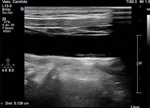
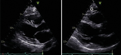
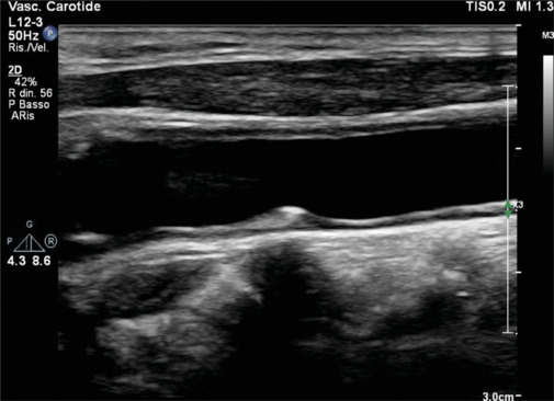
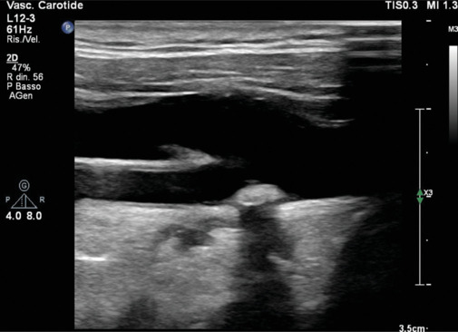
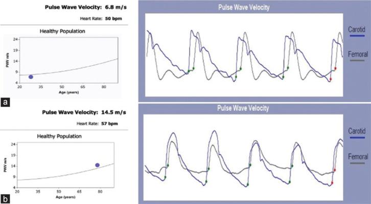
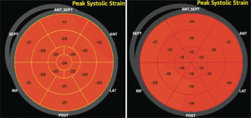
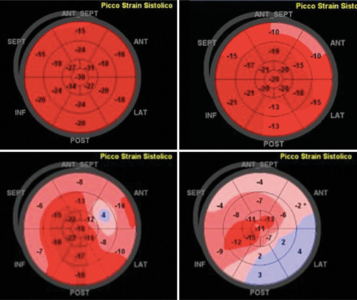
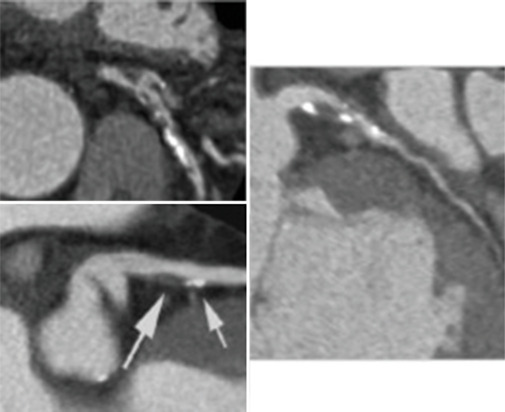
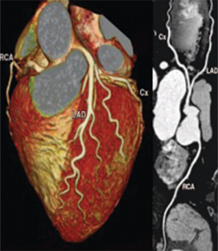
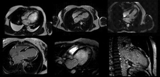
Cardiovascular diseases (CVDs) remain the leading cause of morbidity and mortality worldwide, and traditional preventive measures focus on lifestyle modifications, pharmacologic interventions, and risk stratification. Recently, imaging has emerged as an interesting tool in cardiovascular prevention. This review explores the role of various imaging modalities in early detection, risk assessment, and disease monitoring. Noninvasive techniques such as carotid ultrasound, arterial stiffness assessment, echocardiography, and coronary artery calcium scoring enable the identification of subclinical atherosclerosis and ventricular dysfunction, providing insights that complement conventional risk factors. Coronary computed tomography angiography and cardiac magnetic resonance offer high-resolution visualization of vascular and myocardial pathology, contributing to refined risk stratification. Furthermore, emerging markers such as epicardial adipose tissue and hepatic steatosis are gaining recognition as potential predictors of cardiovascular risk. Advancements in artificial intelligence (AI) are revolutionizing cardiovascular imaging by enhancing image interpretation, automating risk prediction, and facilitating personalized medicine. Future research should focus on optimizing the integration of imaging into clinical workflows, improving risk prediction models, and exploring AI-driven innovations. By exploiting imaging technologies, clinicians could enhance primary and secondary prevention strategies, ultimately reducing the global burden of CVDs.
心血管疾病(CVDs)仍然是全球发病和死亡的主要原因,传统的预防措施集中在生活方式改变、药物干预和风险分层上。最近,影像学已成为心血管预防领域一项引人关注的工具。本综述探讨了各种影像学检查方法在早期检测、风险评估和疾病监测中的作用。诸如颈动脉超声、动脉僵硬度评估、超声心动图和冠状动脉钙化积分等非侵入性技术能够识别亚临床动脉粥样硬化和心室功能障碍,提供补充传统危险因素的见解。冠状动脉计算机断层扫描血管造影和心脏磁共振成像能够对血管和心肌病变进行高分辨率可视化,有助于更精确的风险分层。此外,诸如心外膜脂肪组织和肝脂肪变性等新兴标志物正逐渐被认可为心血管风险的潜在预测指标。人工智能(AI)的进步正在通过增强图像解读、自动化风险预测和推动个性化医疗来彻底改变心血管成像。未来的研究应聚焦于优化影像学在临床工作流程中的整合、改进风险预测模型以及探索人工智能驱动的创新。通过利用成像技术,临床医生可以加强一级和二级预防策略,最终减轻全球心血管疾病负担。