Department of Obstetrics and Gynecology, Radboud University Medical Center, PO Box 9101, 6500 HB Nijmegen, The Netherlands.
Department of Human Genetics, Radboud University Medical Center, PO Box 9101, 6500 HB Nijmegen, The Netherlands.
Hum Reprod. 2019 Sep 29;34(9):1686-1696. doi: 10.1093/humrep/dez135.
What is the X chromosomal content of oocytes and granulosa cells of primordial/primary (small) follicles and stromal cells in ovaries of young patients with Turner's syndrome (TS)?
Small ovarian follicles were detected in one-half of the patients studied, and X chromosome analysis revealed that most oocytes were normal, granulosa cells were largely monosomic, while stromal cells showed a high level of mosaicism.
Most women with TS experience a premature reduction or complete loss of fertility due to an accelerated loss of gametes. To determine whether fertility preservation in this group of patients is feasible, there is a strong need for information on the X chromosomal content of ovarian follicular and stromal cells.
STUDY DESIGN, SIZE, DURATION: Small follicles (<50 μm) and stromal cells were isolated from ovarian tissue of young TS patients and analysed for their X chromosomal content. In addition to ovarian cells, several other cell types from the same patients were analysed.
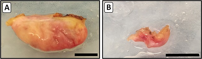
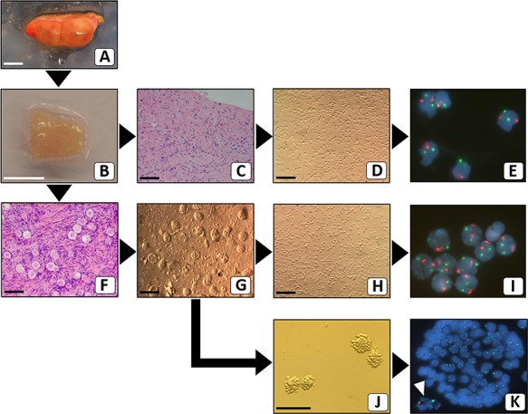
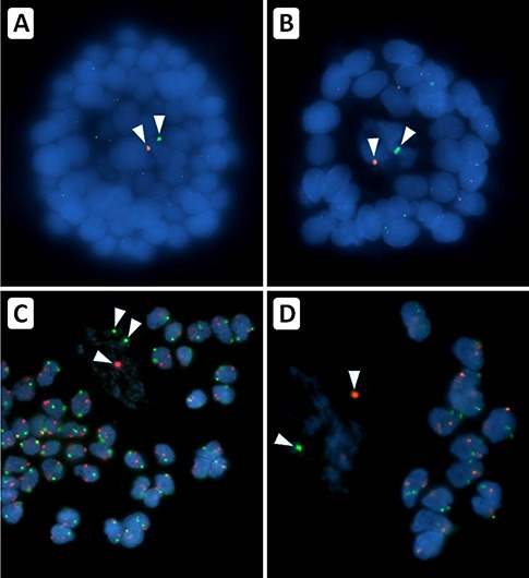
PARTICIPANTS/MATERIALS, SETTING, METHODS: After unilateral ovariectomy, ovarian cortex tissue was obtained from 10 TS patients (aged 2-18 years) with numerical abnormalities of the X chromosome. Ovarian cortex fragments were prepared and cryopreserved. One fragment from each patient was thawed and enzymatically digested to obtain stromal cells and primordial/primary follicles. Stromal cells, granulosa cells and oocytes were analysed by FISH using an X chromosome-specific probe. Extra-ovarian cells (lymphocytes, buccal cells and urine cells) of the same patients were also analysed by FISH. Ovarian tissue used as control was obtained from individuals undergoing oophorectomy as part of their gender affirming surgery.
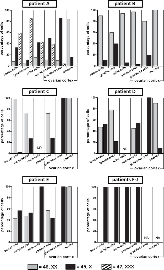
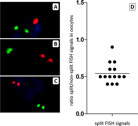
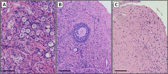
Ovarian follicles were detected in 5 of the 10 patients studied. A method was developed to determine the X chromosomal content of meiosis I arrested oocytes from small follicles. This revealed that 42 of the 46 oocytes (91%) that were analysed had a normal X chromosomal content. Granulosa cells were largely 45,X but showed different levels of X chromosome mosaicism between patients and between follicles of the same patient. Despite the presence of a low percentage (10-45%) of 46,XX ovarian cortex stromal cells, normal macroscopic ovarian morphology was observed. The level of mosaicism in lymphocytes, buccal cells or urine-derived cells was not predictive for mosaicism in ovarian cells.
LIMITATIONS, REASONS FOR CAUTION: The results are based on a small number (n = 5) of TS patient samples but provide evidence that the majority of oocytes have a normal X chromosomal content and that follicles from the same patient can differ with respect to the level of mosaicism of their granulosa cells. The functional consequences of these observations require further investigation.
The results indicate that despite normal ovarian and follicular morphology, stromal cells and granulosa cells of small follicles in patients with TS may display a high level of mosaicism. Furthermore, the level of mosaicism in ovarian cells cannot be predicted from the analysis of extra-ovarian tissue. These findings should be considered by physicians when offering cryopreservation of ovarian tissue as an option for fertility preservation in young TS patients.
STUDY FUNDING/COMPETING INTEREST(S): Unconditional funding was received from Merck B.V. The Netherlands (Number A16-1395) and the foundation 'Radboud Oncologie Fonds' (Number KUN 00007682). The authors have no conflicts of interest.
NCT03381300.
特纳综合征(TS)年轻患者的原始/初级(小)卵泡的卵母细胞和颗粒细胞以及卵巢基质细胞的 X 染色体含量是多少?
在研究的一半患者中检测到小的卵巢卵泡,X 染色体分析显示大多数卵母细胞正常,颗粒细胞主要为单体性,而基质细胞表现出高水平的嵌合性。
大多数 TS 女性由于配子的加速丢失而经历过早或完全丧失生育能力。为了确定在这组患者中是否可行进行生育力保存,需要了解卵巢卵泡和基质细胞的 X 染色体含量的相关信息。
研究设计、大小、持续时间:从小 TS 患者的卵巢组织中分离小卵泡(<50μm)和基质细胞,并分析其 X 染色体含量。除了卵巢细胞外,还分析了同一患者的其他几种细胞类型。
参与者/材料、设置、方法:在单侧卵巢切除术之后,从小 TS 患者(年龄 2-18 岁)的卵巢皮质组织中获得具有 X 染色体数量异常的组织。制备卵巢皮质组织片段并进行冷冻保存。从每位患者中取出一个片段进行解冻,并通过酶消化获得基质细胞和原始/初级卵泡。使用 X 染色体特异性探针通过 FISH 分析基质细胞、颗粒细胞和卵母细胞。同一患者的额外卵巢细胞(淋巴细胞、口腔细胞和尿液细胞)也通过 FISH 进行分析。作为对照的卵巢组织取自作为其性别肯定手术一部分接受卵巢切除术的个体。
在研究的 10 名患者中,有 5 名患者检测到卵巢卵泡。开发了一种方法来确定从小卵泡中检测到的减数分裂 I 阻滞卵母细胞的 X 染色体含量。这表明分析的 46 个卵母细胞中有 42 个(91%)具有正常的 X 染色体含量。颗粒细胞主要为 45,X,但在患者之间和同一患者的卵泡之间表现出不同水平的 X 染色体嵌合性。尽管存在低百分比(10-45%)的 46,XX 卵巢皮质基质细胞,但观察到宏观正常的卵巢形态。淋巴细胞、口腔细胞或尿液衍生细胞中的嵌合率并不能预测卵巢细胞中的嵌合率。
局限性、谨慎的原因:结果基于少数(n=5)TS 患者样本,但提供了证据表明大多数卵母细胞具有正常的 X 染色体含量,并且同一患者的卵泡在其颗粒细胞的嵌合水平方面可能存在差异。这些观察结果的功能后果需要进一步研究。
结果表明,尽管卵巢和卵泡形态正常,但 TS 患者的小卵泡的基质细胞和颗粒细胞可能表现出高水平的嵌合性。此外,卵巢细胞中的嵌合率不能通过分析卵巢外组织来预测。当为年轻 TS 患者提供卵巢组织冷冻保存作为生育力保存的选择时,医生应考虑这些发现。
研究资金/利益冲突:默克公司(荷兰)(编号 A16-1395)和“Radboud 肿瘤学基金”无条件提供资金(编号 KUN 00007682)。作者没有利益冲突。
NCT03381300。