University of Utah Huntsman Cancer Institute, Salt Lake City, UT, USA.
Minnesota Oncology and Virginia Piper Cancer Institute, Fridley, MN, USA.
EBioMedicine. 2019 Sep;47:89-97. doi: 10.1016/j.ebiom.2019.07.066. Epub 2019 Aug 10.
Talimogene laherparepvec (T-VEC) is an intralesionally delivered, modified herpes simplex virus type-1 oncolytic immunotherapy. The biodistribution, shedding, and potential transmission of T-VEC was systematically evaluated during and after completion of therapy in adults with advanced melanoma.
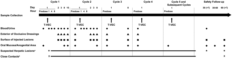
In this phase 2, single-arm, open-label study, T-VEC was administered into injectable lesions initially at 10 plaque-forming units (PFU)/mL, 10 PFU/mL 21 days later, and 10 PFU/mL every 14 (±3) days thereafter. Injected lesions were covered with occlusive dressings for ≥1 week. Blood, urine, and swabs from exterior of occlusive dressings, surface of injected lesions, oral mucosa, anogenital area, and suspected herpetic lesions were collected throughout the study. Detectable T-VEC DNA was determined for each sample type; infectivity was determined for all swabs with detectable T-VEC DNA.
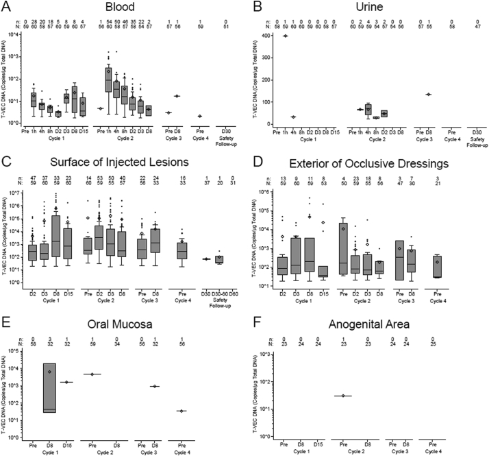
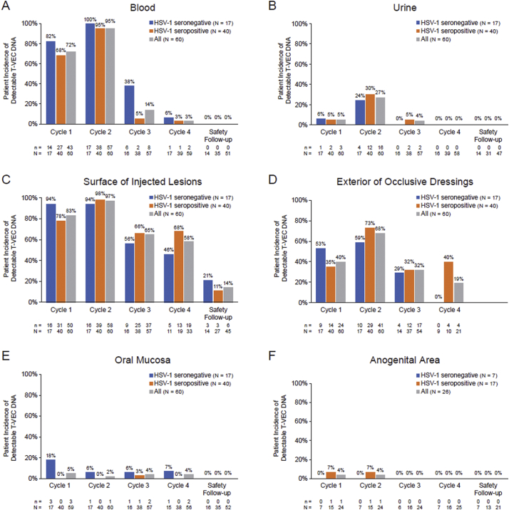
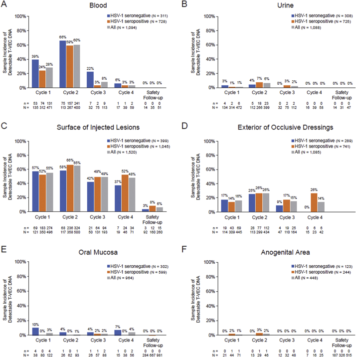
Sixty patients received ≥1 dose of T-VEC. During cycles 1-4, T-VEC DNA was detected in blood (98·3% of patients, 36·7% of samples), urine (31·7% of patients, 3·0% of samples) and swabs from injected lesions (100% of patients, 57·6% of samples), exterior of dressings (80% of patients,19·5% of samples), oral mucosa (8·3% of patients, 2·5% of samples), and anogenital area (8·0% of patients, 1·1% of samples). During the safety follow-up period, T-VEC DNA was only detected on swabs from injected lesions (14% of patients, 5.8% of samples). T-VEC DNA was detected in 4/37 swabs (3/19 patients) of suspected herpetic lesions. Among all samples, only those from the surface of injected lesions tested positive for infectivity (8/740 [1·1%]). Three close contacts reported signs and symptoms of suspected herpetic origin; however, no lesions had detectable T-VEC DNA.
Using current guidelines, T-VEC can be administered safely to patients with advanced melanoma and is unlikely to be transmitted to close contacts with appropriate use of occlusive dressings. FUND: This study was funded by Amgen Inc.: ClinicalTrials.gov, NCT02014441.
替莫唑胺拉帕替尼(T-VEC)是一种瘤内递送的、经过修饰的单纯疱疹病毒 1 型溶瘤免疫疗法。在晚期黑色素瘤成人患者中,在完成治疗期间和之后,系统地评估了 T-VEC 的体内分布、脱落和潜在传播。
在这项 2 期、单臂、开放标签研究中,T-VEC 最初以 10 噬菌斑形成单位(PFU)/mL 的剂量注入可注射病灶,21 天后以 10 PFU/mL 的剂量注入,此后每 14(±3)天以 10 PFU/mL 的剂量注入。注入病灶用密闭敷料覆盖至少 1 周。在整个研究过程中,从密闭敷料的外部、注射病灶的表面、口腔黏膜、肛门生殖器区域和疑似疱疹病灶收集血液、尿液和拭子。对每种样本类型进行可检测 T-VEC DNA 的检测;对所有具有可检测 T-VEC DNA 的拭子进行传染性检测。
60 名患者接受了至少 1 剂 T-VEC。在第 1-4 周期中,血液(98.3%的患者,36.7%的样本)、尿液(31.7%的患者,3.0%的样本)和注射病灶拭子(100%的患者,57.6%的样本)、敷料外部(80%的患者,19.5%的样本)、口腔黏膜(8.3%的患者,2.5%的样本)和肛门生殖器区域(8.0%的患者,1.1%的样本)均检测到 T-VEC DNA。在安全性随访期间,仅在注射病灶拭子上检测到 T-VEC DNA(14%的患者,5.8%的样本)。在 37 份疑似疱疹病灶拭子中,有 4 份(19 名患者中的 3 名)检测到 T-VEC DNA。在所有样本中,只有注射病灶表面的样本检测到具有传染性(740 份样本中的 8 份,1.1%)。3 名密切接触者报告有疑似疱疹起源的体征和症状;然而,没有病变检测到可检测的 T-VEC DNA。
使用当前的指南,替莫唑胺拉帕替尼可安全地给予晚期黑色素瘤患者,并且在适当使用密闭敷料的情况下不太可能传播给密切接触者。
本研究由安进公司资助:ClinicalTrials.gov,NCT02014441。