Liu Qianqian, Li Zhenzhen, Shang Hao, Zhang Qiaochu, Wang Xiaomeng, Zhang Yangyang, Wang Yang, Li Qianru, Li Chunsen, Liu Chunxia, Li Feng
Shihezi University, Shihezi 832002, Xinjiang, P.R. China. a. Department of Pathology, School of Medicine. b. The Key Laboratories for Xinjiang Endemic and Ethnic Diseases, Chinese Ministry of Education. c. The First Affiliated Hospital, School of Medicine.
Department of Pathology, Beijing Chaoyang Hospital, Capital Medical University, Beijing 100020, P. R. China.
J Cancer. 2019 Jul 10;10(18):4326-4332. doi: 10.7150/jca.31730. eCollection 2019.
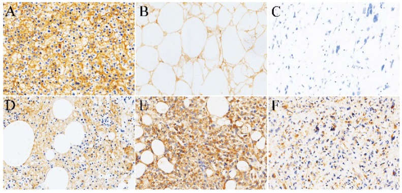
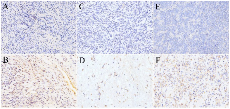
: Soft tissue sarcomas include multiple histological subtypes and are highly aggressive. Moreover, SR-B1 is associated with malignant behavior and poor prognosis in a variety of cancers. However, there have been no attempts to assess whether SR-B1 expression in soft tissue sarcoma. We aimed to detect the expression levels of the SR-B1 protein in soft tissue sarcoma. : We assessed SR-B1 expression via immunohistochemistry and tissue microarrays in 107 soft tissue sarcomas with 4 phenotypes: 26 liposarcomas, 18 Ewing's sarcomas, 20 rhabdomyosarcomas and 43 leiomyosarcomas. : Tumor cell SR-B1 expression was seen in 18/26 (69.2%) liposarcomas, 1/18 (5.55%) Ewing's sarcomas, 1/20 (5.00%) rhabdomyosarcomas, 2/43 (4.70%) leiomyosarcomas and was stained in the cell membrane. We found that SR-B1 expression in liposarcomas (18/26) was significantly higher than that in non-lipomatous sarcomas (4/77) (2 = 49.811, = 0.000) and in well-differentiated liposarcoma (13/15) was significantly higher than that in dedifferentiated liposarcoma (5/11) ( = 0.038). No significant correlation was found between SR-B1 and gender, nationality, size and tumor location ( > 0.05), but it was significantly associated with ages (2 = 11.426, = 0.001) and sarcoma phenotypes (2 = 49.817, = 0.000). : Our findings highlight the highly expression of SR-B1 in liposarcomas. SR-B1 may be a potential biomarker for the diagnosis of liposarcoma and may indicate the degree of differentiation of liposarcomas.
软组织肉瘤包括多种组织学亚型,且具有高度侵袭性。此外,SR-B1与多种癌症的恶性行为和不良预后相关。然而,此前尚未有人尝试评估软组织肉瘤中SR-B1的表达情况。我们旨在检测软组织肉瘤中SR-B1蛋白的表达水平。:我们通过免疫组织化学和组织微阵列评估了107例具有4种表型的软组织肉瘤中SR-B1的表达:26例脂肪肉瘤、18例尤因肉瘤、20例横纹肌肉瘤和43例平滑肌肉瘤。:在18/26(69.2%)的脂肪肉瘤、1/18(5.55%)的尤因肉瘤、1/20(5.00%)的横纹肌肉瘤、2/43(4.70%)的平滑肌肉瘤中可见肿瘤细胞SR-B1表达,且其在细胞膜上呈染色阳性。我们发现脂肪肉瘤(18/26)中的SR-B1表达显著高于非脂肪瘤性肉瘤(4/77)(χ² = 49.811,P = 0.000),且高分化脂肪肉瘤(13/15)中的SR-B1表达显著高于去分化脂肪肉瘤(5/11)(P = 0.038)。未发现SR-B1与性别、国籍、肿瘤大小及肿瘤位置之间存在显著相关性(P > 0.05),但它与年龄(χ² = 11.426,P = 0.001)和肉瘤表型(χ² = 49.817,P = 0.000)显著相关。:我们的研究结果突出了SR-B1在脂肪肉瘤中的高表达。SR-B1可能是诊断脂肪肉瘤的潜在生物标志物,且可能指示脂肪肉瘤的分化程度。