Sukhanova Alyona, Poly Simon, Bozrova Svetlana, Lambert Éléonore, Ewald Maxime, Karaulov Alexander, Molinari Michael, Nabiev Igor
Laboratoire de Recherche en Nanosciences, LRN-EA4682, UFR de Pharmacie, Université de Reims Champagne-Ardenne, Reims, France.
Laboratory of Nano-Bioengineering, Moscow Engineering Physics Institute, National Research Nuclear University MEPhI, Moscow, Russia.
Front Chem. 2019 Jul 30;7:480. doi: 10.3389/fchem.2019.00480. eCollection 2019.
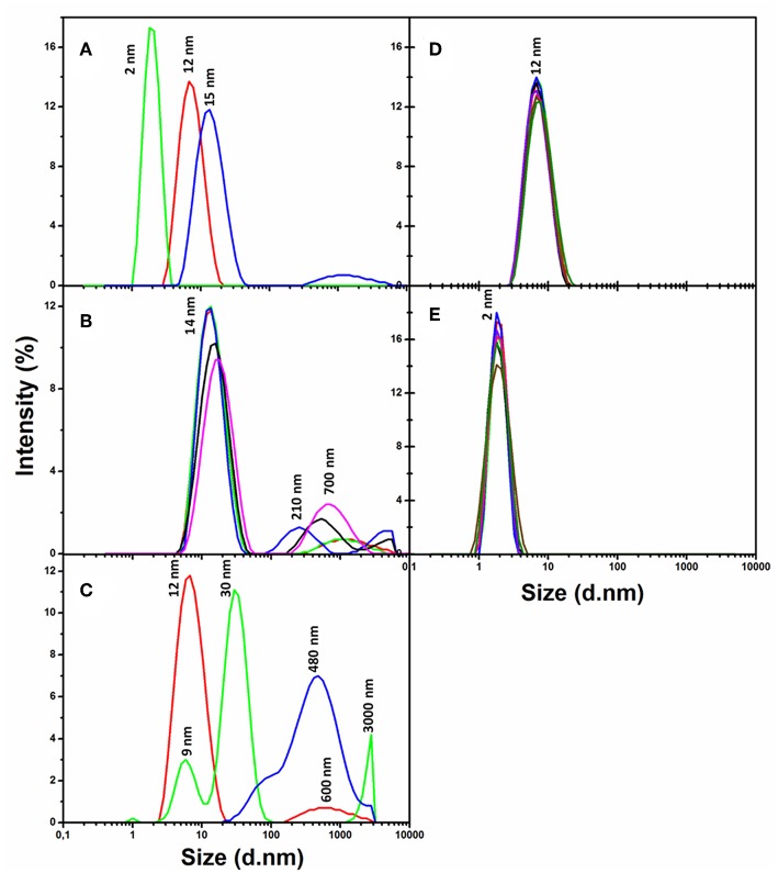
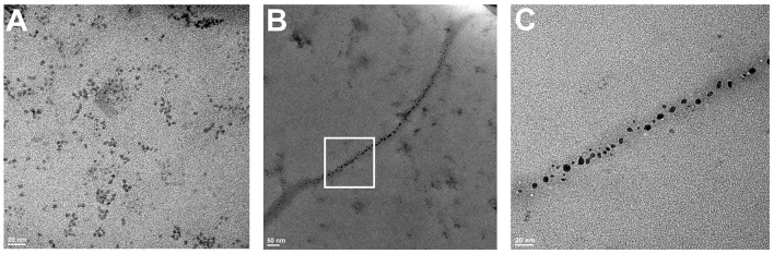
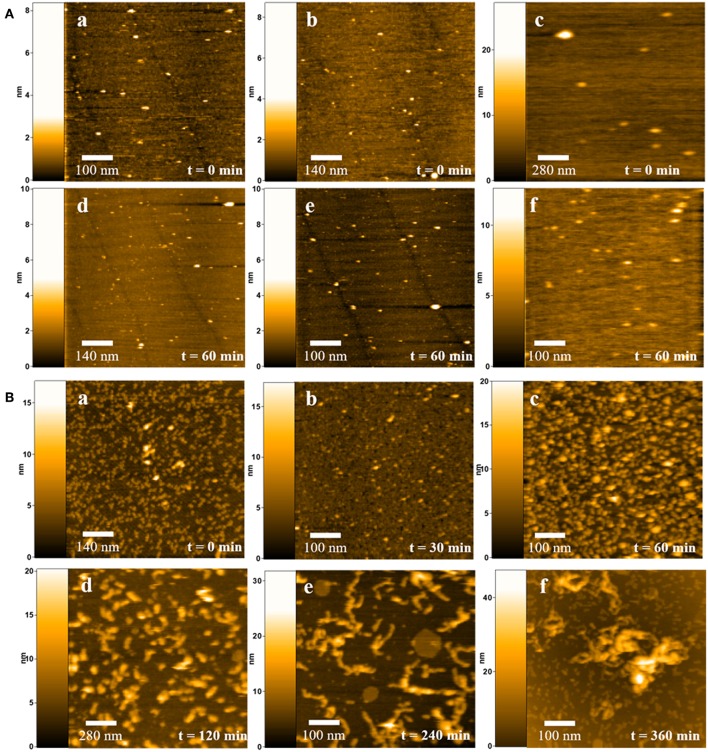
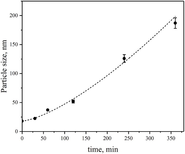
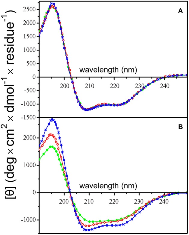
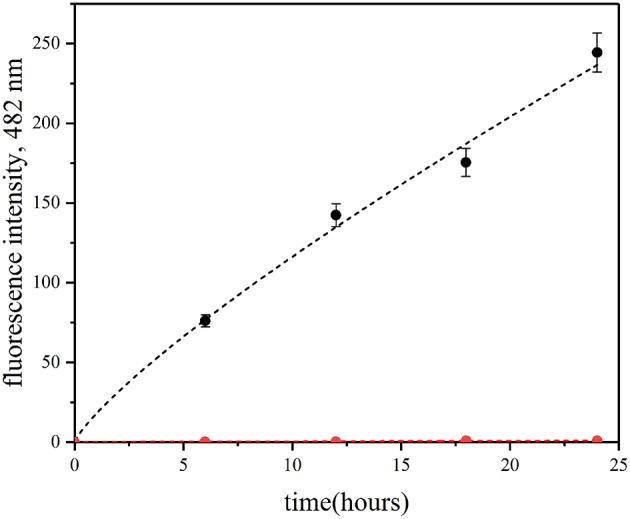
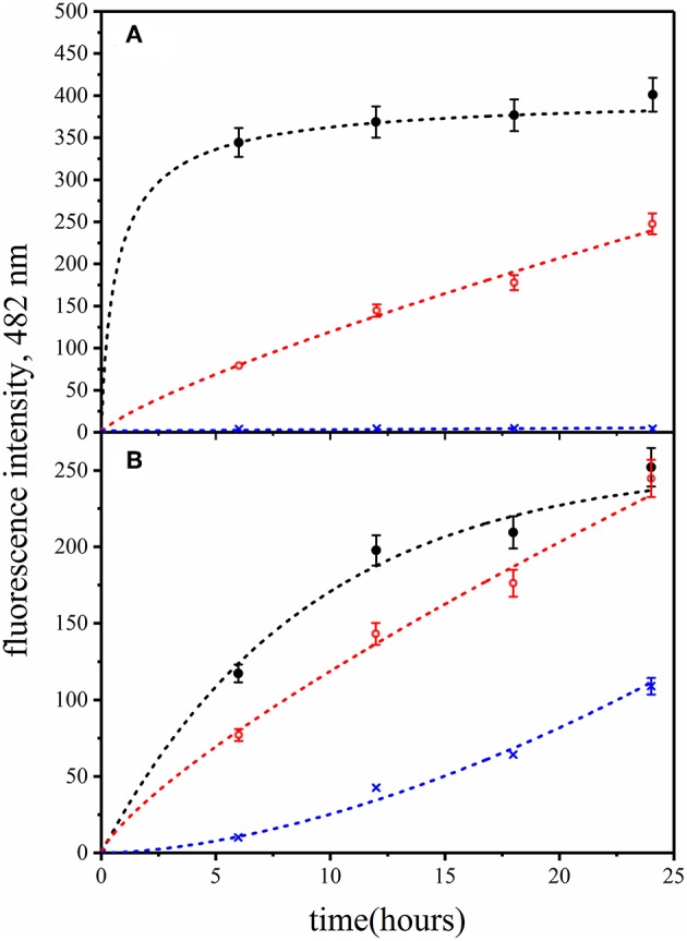
Nanoparticles attract much interest as fluorescent labels for diagnostic and therapeutic tools, although their applications are often hindered by size- and shape-dependent cytotoxicity. This cytotoxicity is related not only to the leak of toxic metals from nanoparticles into a biological solution, but also to molecular cytotoxicity effects determined by the formation of a protein corona, appearance of an altered protein conformation leading to exposure of cryptic epitopes and cooperative effects involved in the interaction of proteins and peptides with nanoparticles. In the last case, nanoparticles may serve, depending on their nature, as centers of self-association or fibrillation of proteins and peptides, provoking amyloid-like proteinopathies, or as inhibitors of self-association of proteins, or they can self-assemble on biopolymers as on templates. In this study, human insulin protein was used to analyze nanoparticle-induced proteinopathy in physiological conditions. It is known that human insulin may form amyloid fibers, but only under extreme experimental conditions (very low pH and high temperatures). Here, we have shown that the quantum dots (QDs) may induce amyloid-like fibrillation of human insulin under physiological conditions through a complex process strongly dependent on the size and surface charge of QDs. The insulin molecular structure and fibril morphology have been shown to be modified at different stages of its fibrillation, which has been proved by comparative analysis of the data obtained using circular dichroism, dynamic light scattering, amyloid-specific thioflavin T (ThT) assay, transmission electron microscopy, and high-speed atomic force microscopy. We have found important roles of the QD size and surface charge in the destabilization of the insulin structure and the subsequent fibrillation. Remodeling of the insulin secondary structure accompanied by remarkable increase in the rate of formation of amyloid-like fibrils under physiologically normal conditions was observed when the protein was incubated with QDs of exact specific diameter coated with slightly negative specific polyethylene glycol (PEG) derivatives. Strongly negatively or slightly positively charged PEG-modified QDs of the same specific diameter or QDs of bigger or smaller diameters had no effect on insulin fibrillation. The observed effects pave the way to the control of amyloidosis proteinopathy by varying the nanoparticle size and surface charge.
纳米颗粒作为诊断和治疗工具的荧光标记物备受关注,尽管其应用常常受到尺寸和形状依赖性细胞毒性的阻碍。这种细胞毒性不仅与有毒金属从纳米颗粒泄漏到生物溶液中有关,还与由蛋白质冠层的形成、导致隐蔽表位暴露的蛋白质构象改变以及蛋白质和肽与纳米颗粒相互作用中涉及的协同效应所决定的分子细胞毒性效应有关。在最后一种情况下,纳米颗粒根据其性质,可能作为蛋白质和肽的自缔合或纤维化中心,引发淀粉样蛋白病,或者作为蛋白质自缔合的抑制剂,或者它们可以在生物聚合物上作为模板进行自组装。在本研究中,使用人胰岛素蛋白来分析生理条件下纳米颗粒诱导的蛋白病。已知人胰岛素可能形成淀粉样纤维,但仅在极端实验条件下(极低pH值和高温)。在这里,我们表明量子点(QDs)在生理条件下可能通过一个强烈依赖于量子点尺寸和表面电荷的复杂过程诱导人胰岛素的淀粉样纤维化。胰岛素分子结构和纤维形态在其纤维化的不同阶段被证明发生了改变,这通过使用圆二色性、动态光散射、淀粉样特异性硫黄素T(ThT)测定、透射电子显微镜和高速原子力显微镜获得的数据的比较分析得到了证实。我们发现量子点尺寸和表面电荷在胰岛素结构的不稳定和随后的纤维化中起着重要作用。当蛋白质与涂有略带负电荷的特定聚乙二醇(PEG)衍生物的精确特定直径的量子点孵育时,观察到胰岛素二级结构的重塑伴随着在生理正常条件下淀粉样纤维形成速率的显著增加。相同特定直径的强负电荷或略带正电荷的PEG修饰量子点或更大或更小直径的量子点对胰岛素纤维化没有影响。观察到的这些效应为通过改变纳米颗粒尺寸和表面电荷来控制淀粉样蛋白病铺平了道路。