Translational Molecular Imaging Group, Max Planck Institute for Experimental Medicine, Göttingen, Germany.
Laboratoire de Recherche en Nanosciences (LRN-EA4682), Université de Reims Champagne-Ardenne, 51100, Reims, France.
Sci Rep. 2018 Mar 15;8(1):4595. doi: 10.1038/s41598-018-22973-8.
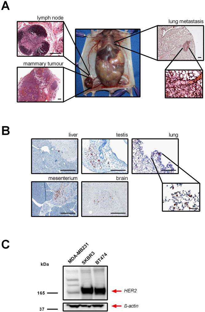
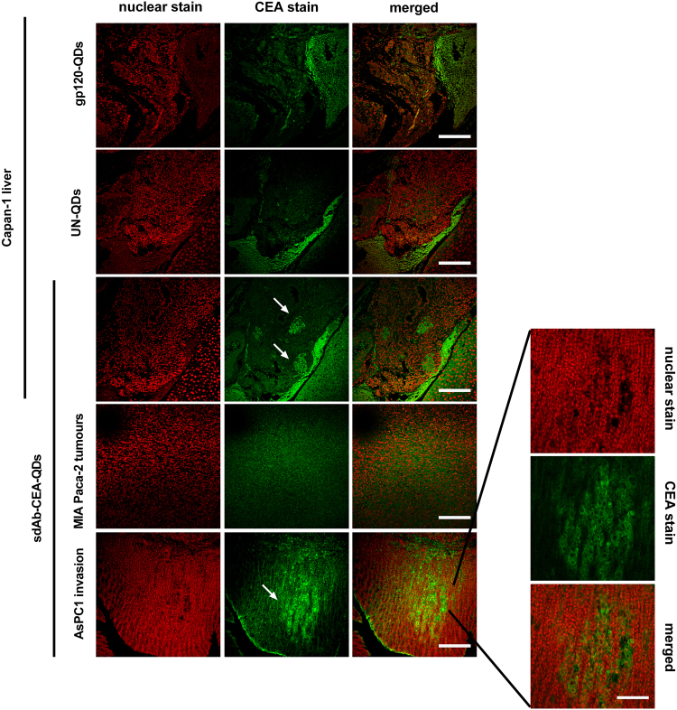
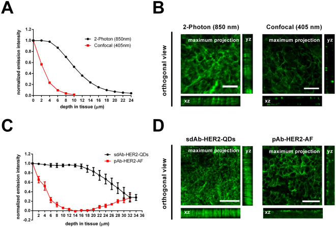
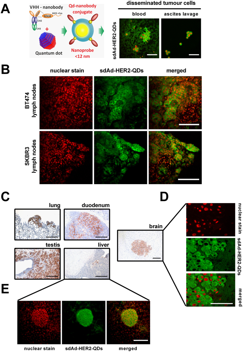
Early detection of malignant tumours and, especially, micrometastases and disseminated tumour cells is still a challenge. In order to implement highly sensitive diagnostic tools we demonstrate the use of nanoprobes engineered from nanobodies (single-domain antibodies, sdAbs) and fluorescent quantum dots (QDs) for single- and two-photon detection and imaging of human micrometastases and disseminated tumour cells in ex vivo biological samples of breast and pancreatic metastatic tumour mouse models expressing human epidermal growth factor receptor 2 (HER2) or carcinoembryonic antigen (CEA). By staining thin (5-10 µm) paraffin and thick (50 µm) agarose tissue sections, we detected HER2- and CEA-positive human tumour cells infiltrating the surrounding tissues or metastasizing to different organs, including the brain, testis, lung, liver, and lymph nodes. Compared to conventional fluorescently labelled antibodies the sdAb-HER2-QD and sdAb-CEA-QD nanoprobes are superior in detecting micrometastases in tissue sections by lower photobleaching and higher brightness of fluorescence signals ensuring much better discrimination of positive signals versus background. Very high two-photon absorption cross-sections of QDs and small size of the nanoprobes ensure efficient imaging of thick tissue sections unattainable with conventional fluorescent probes. The nanobody-QD probes will help to improve early cancer diagnosis and prognosis of progression by assessing metastasis.
早期检测恶性肿瘤,尤其是微转移和播散的肿瘤细胞仍然是一个挑战。为了实现高灵敏度的诊断工具,我们展示了使用纳米体(单域抗体,sdAb)和荧光量子点(QDs)工程化的纳米探针,用于单光子和双光子检测和成像人微转移和播散的肿瘤细胞在表达人表皮生长因子受体 2(HER2)或癌胚抗原(CEA)的乳腺癌和胰腺癌转移肿瘤小鼠模型的离体生物样本中。通过对 5-10μm 厚的石蜡和 50μm 厚的琼脂糖组织切片进行染色,我们检测到浸润周围组织或转移到不同器官(包括脑、睾丸、肺、肝和淋巴结)的 HER2 和 CEA 阳性人肿瘤细胞。与传统荧光标记抗体相比,sdAb-HER2-QD 和 sdAb-CEA-QD 纳米探针在组织切片中检测微转移时具有更低的光漂白和更高的荧光信号亮度,从而能够更好地区分阳性信号与背景。量子点的高双光子吸收截面和纳米探针的小尺寸确保了对传统荧光探针无法成像的厚组织切片的高效成像。这些纳米体-QD 探针将有助于通过评估转移来提高癌症的早期诊断和进展预后。