Department of Pharmacy, Faculty of Health and Medical Sciences, University of Copenhagen, Copenhagen, Denmark.
Department of Biology, Faculty of Science, University of Copenhagen, Copenhagen, Denmark.
PLoS One. 2019 Aug 19;14(8):e0221103. doi: 10.1371/journal.pone.0221103. eCollection 2019.
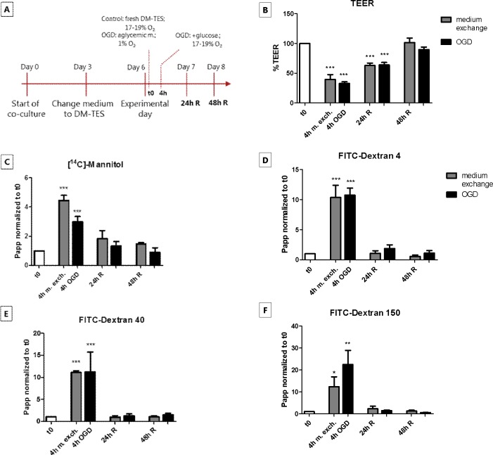
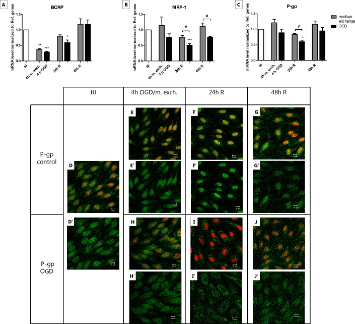
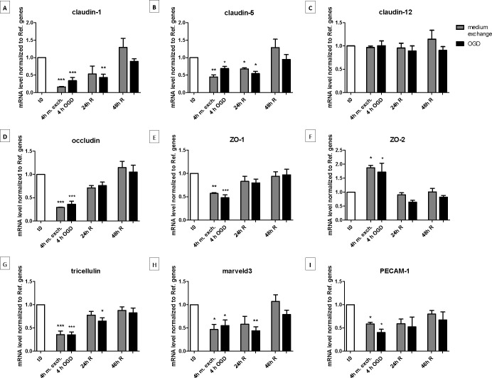
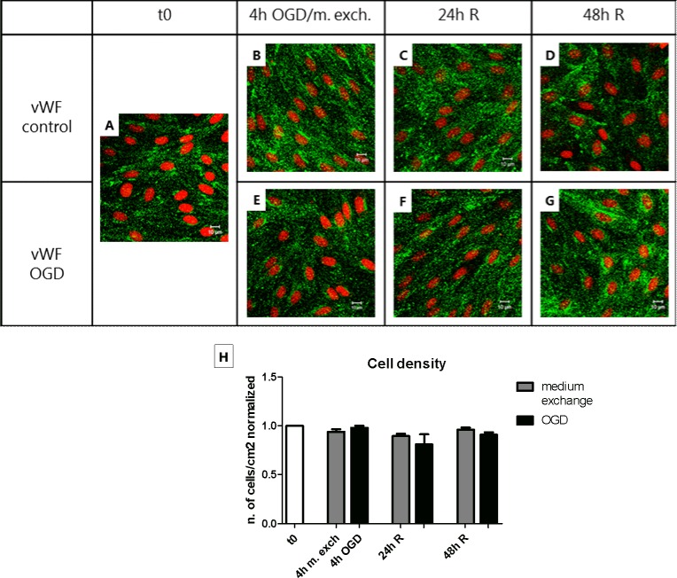
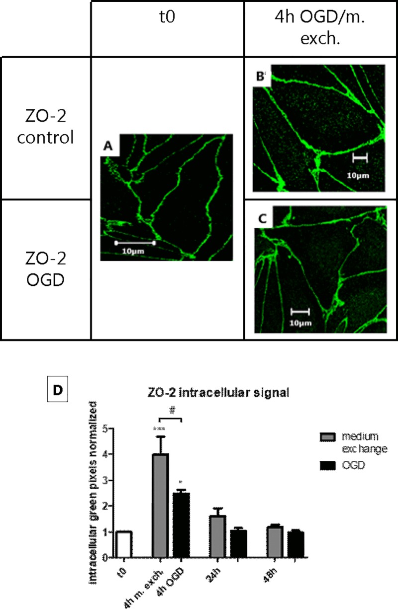
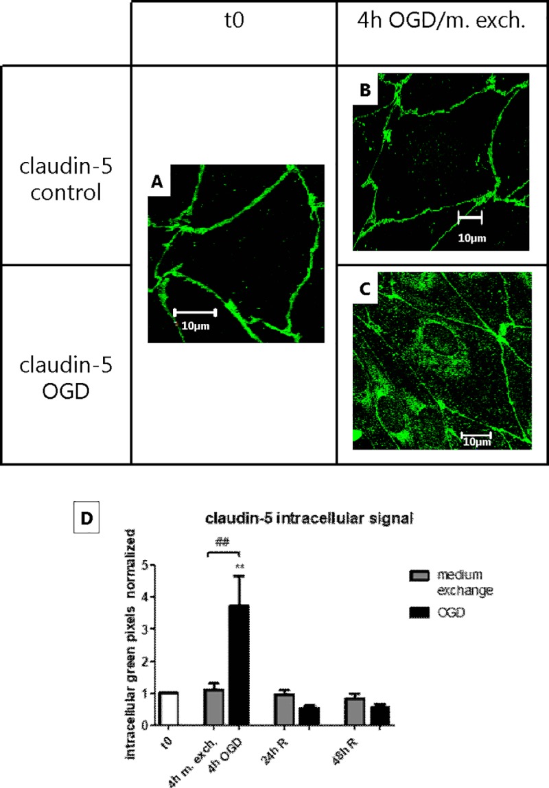
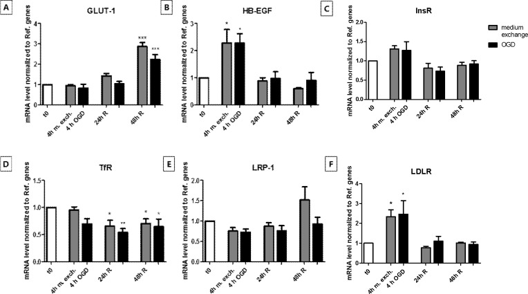
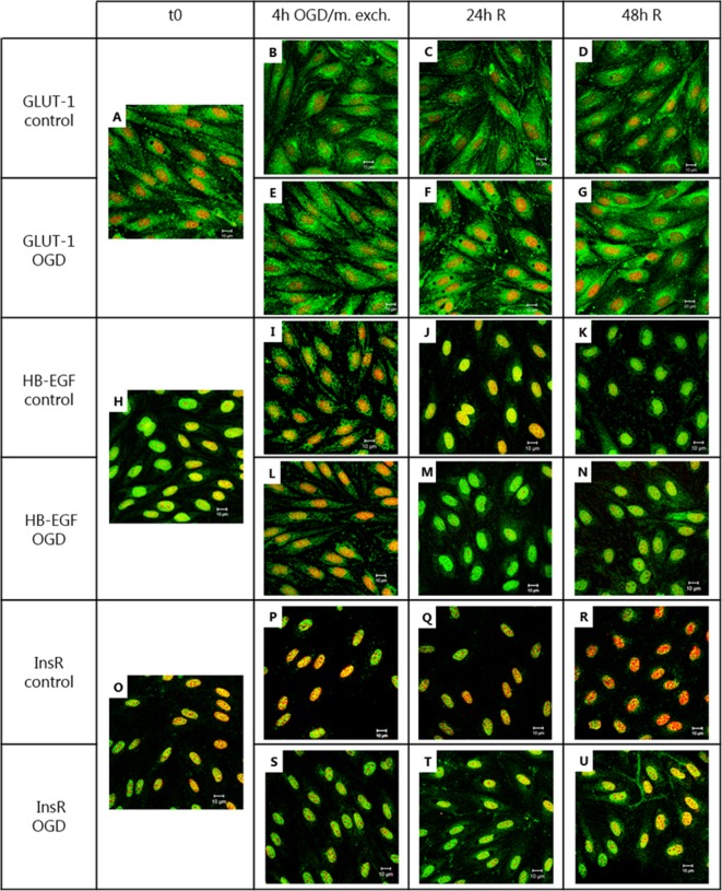
Ischemic stroke has been shown to induce breakdown of the blood-brain barrier, although these changes are not fully characterized. Oxygen-glucose deprivation (OGD) has been used to investigate the effects of ischemia in cultured brain capillary endothelial cells, however this involves a change of medium which in itself may affect the cells. The aim of the present study was to investigate the effect of OGD and simple medium exchange followed by 48 h of reperfusion on barrier properties of primary bovine endothelial cells co-cultured with rat astrocytes. Barrier properties were evaluated by transendothelial electrical resistance measurements, passive permeability of flux markers, RT-qPCR and immunocytochemistry. Both OGD and simple medium exchange caused an increase in endothelial monolayer permeability. This correlated with reduced transcript levels of a number of tight junction and tight junction-associated proteins (claudin-1, claudin-5, occludin, ZO-1, tricellulin, marveld3 and PECAM-1), as well as with altered transcript level of several transporters and receptors (GLUT-1, HB-EGF, InsR, TfR, two members of the low density lipoprotein receptor family, LDLR and LRP-1, and the efflux transporter BCRP). In contrast, effects induced specifically by OGD were transient de-localization of claudin-5 from the junction zone, increased InsR localization at the plasma membrane and transient downregulation of MRP-1 and P-gp transcript levels. In conclusion, OGD caused changes in claudin-5 and InsR localization, as well as in MRP-1 and P-gp transcript levels. Our results however also indicated that medium exchange alone caused changes in functional barrier properties and expression levels of wide range of proteins.
缺血性中风已被证明会导致血脑屏障的破坏,尽管这些变化尚未完全得到阐明。氧葡萄糖剥夺(OGD)已被用于研究培养的脑毛细血管内皮细胞中的缺血影响,但这涉及到培养基的变化,而培养基的变化本身可能会影响细胞。本研究旨在研究 OGD 和简单的培养基交换后再进行 48 小时再灌注对与大鼠星形胶质细胞共培养的原代牛内皮细胞的屏障特性的影响。通过跨内皮电阻测量、通量标记物的被动通透性、RT-qPCR 和免疫细胞化学评估屏障特性。OGD 和简单的培养基交换都会导致内皮单层通透性增加。这与许多紧密连接和紧密连接相关蛋白(claudin-1、claudin-5、occludin、ZO-1、tricellulin、marveld3 和 PECAM-1)的转录水平降低以及几种转运体和受体(GLUT-1、HB-EGF、InsR、TfR、低密度脂蛋白受体家族的两个成员、LDLR 和 LRP-1 以及外排转运蛋白 BCRP)的转录水平改变有关。相比之下,OGD 特异性诱导的作用是 claudin-5 从连接区的去定位、InsR 在质膜上的定位增加以及 MRP-1 和 P-gp 转录水平的短暂下调。总之,OGD 导致 claudin-5 和 InsR 定位以及 MRP-1 和 P-gp 转录水平的改变。然而,我们的结果还表明,仅培养基交换就会引起广泛范围的蛋白质的功能屏障特性和表达水平的改变。