Department of Morphology, Institute of Anatomy and Anthropology, Riga Stradins University, Kronvalda Boulevard 9, LV-1010 Riga, Latvia.
Pauls Stradins Clinical University Hospital, Pilsonu street 13, LV-1002 Riga, Latvia.
Medicina (Kaunas). 2019 Aug 17;55(8):496. doi: 10.3390/medicina55080496.
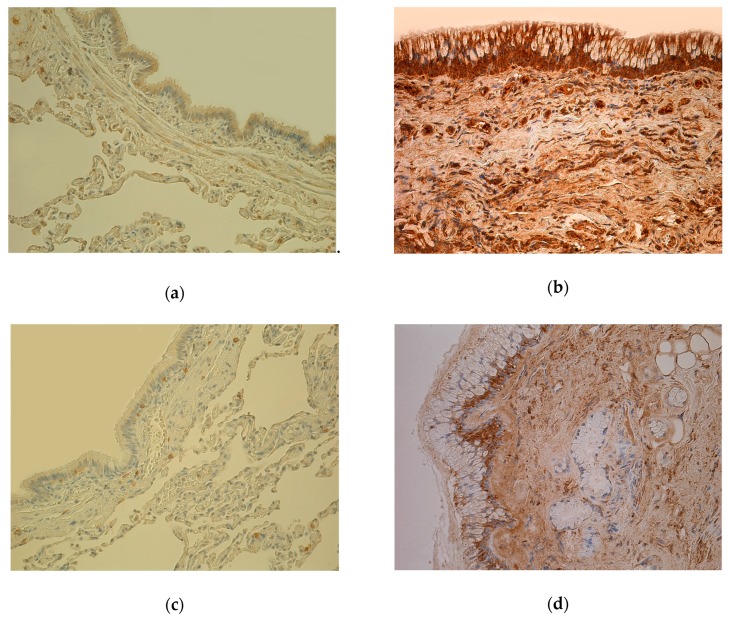
Intercellular signaling networks with high complexity cause a spectrum of mechanisms achieving chronic obstructive pulmonary disease (COPD) that still question many uncertainties. Immunoreactive cells in bronchial tissue obtained from 40 COPD patients and 49 healthy control subjects were detected by biotin-streptavidin immunohistochemistry method for the following markers of IL-1α, IL-4, IL-6, IL-7, IL-8, IL-10, IL-12, TNF-α, MMP-2, TIMP-2, TGF-β1, Hsp-70, hBD-2, hBD-3, hBD-4. Overall the highest numbers (from mostly moderate (++) to abundance (++++)) of IL-1α, IL-4, IL-7, IL-8, IL-10, IL-12, MMP-2, TIMP-2, TGF-β1 immunoreactive cells were marked increasingly in the blood vessel wall, connective tissue, and bronchial epithelium of COPD-affected lung, respectively. We found statistically significant ( < 0.05) higher numbers of immunoreactive cells positive for all of examined interleukins, TNF-α, MMP-2, TIMP-2, TGF-β1, hBD-2, and hBD-3 in the COPD-affected lung compared to the control group, but not for Hsp-70 and hBD-4. COPD-affected lung tissue exhibits mostly inflammatory response patterns of increased IL-1α, IL-4, IL-8, IL-12, and TNF-α, especially in the airway epithelium. Increased MMP-2 and TGF-β1, but decreased Hsp-70, proposes pronounced tissue damage and remodeling in COPD. High numbers of hBD-2 and hBD-3 immunoreactive cells may highlight antimicrobial activity in COPD within stable regulation of local immunity.
细胞间信号网络具有高度复杂性,导致慢性阻塞性肺疾病(COPD)的机制谱仍存在许多不确定性。采用生物素-链霉亲和素免疫组织化学方法检测 40 例 COPD 患者和 49 例健康对照者支气管组织中的免疫反应细胞,用于 IL-1α、IL-4、IL-6、IL-7、IL-8、IL-10、IL-12、TNF-α、MMP-2、TIMP-2、TGF-β1、Hsp-70、hBD-2、hBD-3、hBD-4 的以下标志物。总体而言,IL-1α、IL-4、IL-7、IL-8、IL-10、IL-12、MMP-2、TIMP-2、TGF-β1 免疫反应细胞的数量(大多为中度(++)至丰富(++++))在 COPD 受累肺的血管壁、结缔组织和支气管上皮中分别呈递增趋势。我们发现,与对照组相比,COPD 受累肺中所有检查的白细胞介素、TNF-α、MMP-2、TIMP-2、TGF-β1、hBD-2 和 hBD-3 的免疫反应细胞数量均有统计学意义(<0.05)增加,但 Hsp-70 和 hBD-4 则不然。COPD 受累肺组织表现出以增加的 IL-1α、IL-4、IL-8、IL-12 和 TNF-α为主的炎症反应模式,尤其是在气道上皮中。MMP-2 和 TGF-β1 的增加,而 Hsp-70 的减少,提示 COPD 中存在明显的组织损伤和重塑。大量 hBD-2 和 hBD-3 免疫反应细胞可能突出了 COPD 中局部免疫稳定的抗菌活性。