• 文献检索
  • 文档翻译
  • 深度研究
  • 学术资讯
  • Suppr Zotero 插件Zotero 插件
  • 邀请有礼
  • 套餐&价格
  • 历史记录
应用&插件
Suppr Zotero 插件Zotero 插件浏览器插件Mac 客户端Windows 客户端微信小程序
定价
高级版会员购买积分包购买API积分包
服务
文献检索文档翻译深度研究API 文档MCP 服务
关于我们
关于 Suppr公司介绍联系我们用户协议隐私条款
关注我们

Suppr 超能文献

核心技术专利:CN118964589B侵权必究
粤ICP备2023148730 号-1Suppr @ 2026

文献检索

告别复杂PubMed语法,用中文像聊天一样搜索,搜遍4000万医学文献。AI智能推荐,让科研检索更轻松。

立即免费搜索

文件翻译

保留排版,准确专业,支持PDF/Word/PPT等文件格式,支持 12+语言互译。

免费翻译文档

深度研究

AI帮你快速写综述,25分钟生成高质量综述,智能提取关键信息,辅助科研写作。

立即免费体验

4D 血流磁共振成像衍生的二尖瓣反流定量方法的系统评价

A Systematic Review of 4D-Flow MRI Derived Mitral Regurgitation Quantification Methods.

作者信息

Fidock Benjamin, Barker Natasha, Balasubramanian Nithin, Archer Gareth, Fent Graham, Al-Mohammad Abdullah, Richardson James, O'Toole Laurence, Briffa Norman, Rothman Alexander, van der Geest Rob, Hose Rod, Wild James M, Swift Andrew J, Garg Pankaj

机构信息

Department of Infection, Immunity & Cardiovascular Disease, University of Sheffield, Sheffield, United Kingdom.

Sheffield Teaching Hospitals NHS Foundation Trust, Sheffield, United Kingdom.

出版信息

Front Cardiovasc Med. 2019 Aug 2;6:103. doi: 10.3389/fcvm.2019.00103. eCollection 2019.

DOI:10.3389/fcvm.2019.00103
PMID:31428619
原文链接:https://pmc.ncbi.nlm.nih.gov/articles/PMC6688118/
Abstract

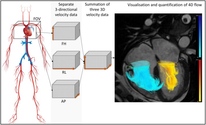
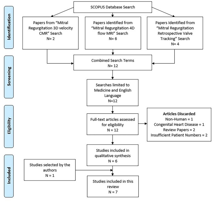
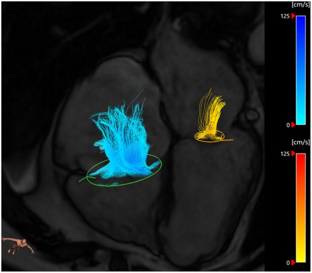
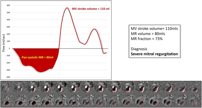
Four-dimensional flow cardiac magnetic resonance (4D flow CMR) is an emerging non-invasive imaging technology that can be used to quantify mitral regurgitation (MR) volume. Current methods of quantification have demonstrated limitations in accurate analysis, particularly in difficult cases such as complex congenital heart disease. 4D flow CMR methods aim to circumvent these limitations and allow accurate quantification of MR volume even in complex cases. This systematic review aims to summarize the available literature on 4D flow CMR MR quantification methods and examine their ability to accurately classify MR severity. Structured searches were carried out on Medline and EMBASE in December 2018 to identify suitable research outcome studies. The titles and abstracts were screened for relevance, with a third adjudicator utilized when study suitability was uncertain. Seven studies met the eligibility criteria and were included in the systematic review. The most widely used 4D flow MRI method was retrospective valve tracking (RVT) which was examined in five papers. The key finding of these papers was that RVT is a reliable and accurate method of regurgitant volume quantification. MR quantification through 4D flow MRI is both feasible and accurate. The evidence gathered suggests that for MR assessment, 4D flow MRI is potentially as accurate and reliable to echocardiography and may be complementary to this technique. Further work on MR quantification 4D flow image analysis is needed to determine the most accurate analysis technique and to demonstrate 4D flow MRI as a predictor of clinical outcome. CRD42019122837, http://www.crd.york.ac.uk/PROSPERO/display_record.php?ID=CRD42019122837.

摘要

四维血流心脏磁共振成像(4D流CMR)是一种新兴的非侵入性成像技术,可用于量化二尖瓣反流(MR)容积。目前的量化方法在准确分析方面存在局限性,尤其是在复杂先天性心脏病等困难病例中。4D流CMR方法旨在克服这些局限性,即使在复杂病例中也能准确量化MR容积。本系统评价旨在总结关于4D流CMR MR量化方法的现有文献,并检验其准确分类MR严重程度的能力。2018年12月在Medline和EMBASE上进行了结构化检索,以确定合适的研究结果研究。对标题和摘要进行相关性筛选,当研究适用性不确定时使用第三位评判员。七项研究符合纳入标准并被纳入系统评价。最广泛使用的4D流MRI方法是回顾性瓣膜追踪(RVT),五篇论文对其进行了研究。这些论文的主要发现是,RVT是一种可靠且准确的反流容积量化方法。通过4D流MRI进行MR量化既可行又准确。收集到的证据表明,对于MR评估,4D流MRI可能与超声心动图一样准确可靠,并且可能是该技术的补充。需要进一步开展关于MR量化4D流图像分析的工作,以确定最准确的分析技术,并证明4D流MRI可作为临床结果的预测指标。CRD42019122837,http://www.crd.york.ac.uk/PROSPERO/display_record.php?ID=CRD42019122837

https://cdn.ncbi.nlm.nih.gov/pmc/blobs/f9bf/6688118/4c6d142dd5d6/fcvm-06-00103-g0007.jpg
https://cdn.ncbi.nlm.nih.gov/pmc/blobs/f9bf/6688118/370c811eae3f/fcvm-06-00103-g0001.jpg
https://cdn.ncbi.nlm.nih.gov/pmc/blobs/f9bf/6688118/a7ea968b19f8/fcvm-06-00103-g0002.jpg
https://cdn.ncbi.nlm.nih.gov/pmc/blobs/f9bf/6688118/53d9ccb8136e/fcvm-06-00103-g0003.jpg
https://cdn.ncbi.nlm.nih.gov/pmc/blobs/f9bf/6688118/0156f32d4e35/fcvm-06-00103-g0004.jpg
https://cdn.ncbi.nlm.nih.gov/pmc/blobs/f9bf/6688118/043c2a1c2bdf/fcvm-06-00103-g0005.jpg
https://cdn.ncbi.nlm.nih.gov/pmc/blobs/f9bf/6688118/62bbd222d996/fcvm-06-00103-g0006.jpg
https://cdn.ncbi.nlm.nih.gov/pmc/blobs/f9bf/6688118/4c6d142dd5d6/fcvm-06-00103-g0007.jpg

相似文献

1
A Systematic Review of 4D-Flow MRI Derived Mitral Regurgitation Quantification Methods.4D 血流磁共振成像衍生的二尖瓣反流定量方法的系统评价
Front Cardiovasc Med. 2019 Aug 2;6:103. doi: 10.3389/fcvm.2019.00103. eCollection 2019.
2
Quantification of regurgitation in mitral valve prolapse with four-dimensional flow cardiovascular magnetic resonance.四维血流心脏磁共振定量评估二尖瓣脱垂伴反流。
J Cardiovasc Magn Reson. 2021 Jul 8;23(1):87. doi: 10.1186/s12968-021-00783-8.
3
Direct mitral regurgitation quantification in hypertrophic cardiomyopathy using 4D flow CMR jet tracking: evaluation in comparison to conventional CMR.应用 4D 流磁共振喷射轨迹法直接定量分析肥厚型心肌病的二尖瓣反流:与传统磁共振比较的评估。
J Cardiovasc Magn Reson. 2021 Dec 6;23(1):138. doi: 10.1186/s12968-021-00828-y.
4
4D flow MRI quantification of mitral and tricuspid regurgitation: Reproducibility and consistency relative to conventional MRI.四维流磁共振成像定量评估二尖瓣和三尖瓣反流:与常规磁共振成像的可重复性和一致性。
J Magn Reson Imaging. 2018 Oct;48(4):1147-1158. doi: 10.1002/jmri.26040. Epub 2018 Apr 11.
5
Grading mitral regurgitation using 4D flow CMR: Comparison to transthoracic echocardiography.使用 4D 流心脏磁共振对二尖瓣反流进行分级:与经胸超声心动图的比较。
Echocardiography. 2022 Jun;39(6):783-793. doi: 10.1111/echo.15364. Epub 2022 May 10.
6
Impact of measurement location on direct mitral regurgitation quantification using four-dimensional flow cardiovascular magnetic resonance.测量位置对使用四维血流心血管磁共振定量二尖瓣反流的影响。
J Cardiovasc Magn Reson. 2025 Jan 26;27(1):101847. doi: 10.1016/j.jocmr.2025.101847.
7
A Systematic Review of Right Ventricular Diastolic Assessment by 4D Flow CMR.4D 流 MRI 右心室舒张功能评估的系统评价
Biomed Res Int. 2019 Mar 14;2019:6074984. doi: 10.1155/2019/6074984. eCollection 2019.
8
Mitral valve regurgitation assessed by intraventricular CMR 4D-flow: a systematic review on the technological aspects and potential clinical applications.通过心室内CMR 4D血流评估二尖瓣反流:关于技术方面和潜在临床应用的系统评价
Int J Cardiovasc Imaging. 2023 Oct;39(10):1963-1977. doi: 10.1007/s10554-023-02893-z. Epub 2023 Jun 16.
9
Flow quantification within the aortic ejection tract using 4D flow cardiac MRI in patients with bicuspid aortic valve: Implications for the assessment of aortic regurgitation.使用4D流心脏磁共振成像对二叶式主动脉瓣患者主动脉射血通道内的血流定量分析:对主动脉瓣反流评估的意义
Diagn Interv Imaging. 2024 Dec;105(12):498-506. doi: 10.1016/j.diii.2024.09.001. Epub 2024 Sep 12.
10
Standard and emerging CMR methods for mitral regurgitation quantification.用于二尖瓣反流定量的标准和新兴 CMR 方法。
Int J Cardiol. 2021 May 15;331:316-321. doi: 10.1016/j.ijcard.2021.01.066. Epub 2021 Feb 3.

引用本文的文献

1
Comprehensive 4D-flow cardiac magnetic resonance evaluation of the descending thoracic aorta in aortic regurgitation.主动脉瓣反流患者胸降主动脉的综合四维血流心脏磁共振评估
Eur Heart J Imaging Methods Pract. 2025 Jan 7;3(1):qyaf002. doi: 10.1093/ehjimp/qyaf002. eCollection 2025 Jan.
2
Cardiovascular magnetic resonance imaging in mitral valve disease.二尖瓣疾病的心血管磁共振成像
Eur Heart J. 2025 Feb 14;46(7):606-619. doi: 10.1093/eurheartj/ehae801.
3
Quadratic stratification of left ventricular hypertrophy and association with mitral insufficiency grading: a retrospective study using cardiac magnetic resonance.

本文引用的文献

1
Differences in Two- and Three-Dimensional Assessment of the Mitral Valve by Novices and Experts, Illustrated Using Anterior Mitral Valve Leaflet Length.新手与专家对二尖瓣进行二维和三维评估的差异,以前二尖瓣叶长度为例说明
J Cardiothorac Vasc Anesth. 2019 Apr;33(4):1022-1028. doi: 10.1053/j.jvca.2018.07.001. Epub 2018 Jul 7.
2
Automated Cardiac Valve Tracking for Flow Quantification with Four-dimensional Flow MRI.基于四维血流磁共振的自动心脏瓣膜追踪技术用于流量量化。
Radiology. 2019 Jan;290(1):70-78. doi: 10.1148/radiol.2018180807. Epub 2018 Oct 30.
3
Advanced Imaging Techniques for Mitral Regurgitation.
左心室肥厚的二次分层及其与二尖瓣关闭不全分级的关联:一项使用心脏磁共振成像的回顾性研究
Cardiovasc Diagn Ther. 2024 Aug 31;14(4):589-608. doi: 10.21037/cdt-23-466. Epub 2024 Aug 6.
4
Nitrogen-15 dynamic nuclear polarization of nicotinamide derivatives in biocompatible solutions.氮-15 动态核极化在生物相容性溶液中烟酰胺衍生物的应用。
Sci Adv. 2023 Aug 25;9(34):eadd3643. doi: 10.1126/sciadv.add3643. Epub 2023 Aug 23.
5
Mitral valve regurgitation assessed by intraventricular CMR 4D-flow: a systematic review on the technological aspects and potential clinical applications.通过心室内CMR 4D血流评估二尖瓣反流:关于技术方面和潜在临床应用的系统评价
Int J Cardiovasc Imaging. 2023 Oct;39(10):1963-1977. doi: 10.1007/s10554-023-02893-z. Epub 2023 Jun 16.
6
Multicenter research priorities in pediatric CMR: results of a collaborative wiki survey.多中心儿科心脏磁共振研究重点:协作式维基调查结果。
Sci Rep. 2023 Jun 3;13(1):9022. doi: 10.1038/s41598-023-34720-9.
7
Echocardiography vs. CMR in the Quantification of Chronic Mitral Regurgitation: A Happy Marriage or Stormy Divorce?超声心动图与心脏磁共振成像在慢性二尖瓣反流定量评估中的比较:是美满结合还是激烈分离?
J Cardiovasc Dev Dis. 2023 Mar 31;10(4):150. doi: 10.3390/jcdd10040150.
8
Automated 4D flow cardiac MRI pipeline to derive peak mitral inflow diastolic velocities using short-axis cine stack: two centre validation study against echocardiographic pulse-wave doppler.使用短轴电影堆栈自动生成 4D 流心脏 MRI 管道以获得峰值二尖瓣流入舒张速度:与超声心动图脉冲波多普勒的双中心验证研究。
BMC Cardiovasc Disord. 2023 Jan 16;23(1):24. doi: 10.1186/s12872-023-03052-x.
9
Kat-ARC accelerated 4D flow CMR: clinical validation for transvalvular flow and peak velocity assessment.Kat-ARC 加速 4D 流心脏磁共振:经瓣口流量和峰值速度评估的临床验证。
Eur Radiol Exp. 2022 Sep 22;6(1):46. doi: 10.1186/s41747-022-00299-5.
10
Rationale and clinical applications of 4D flow cardiovascular magnetic resonance in assessment of valvular heart disease: a comprehensive review.4D 血流心血管磁共振评估心脏瓣膜病的原理及临床应用:全面综述。
J Cardiovasc Magn Reson. 2022 Aug 22;24(1):49. doi: 10.1186/s12968-022-00882-0.
二尖瓣反流的高级影像学技术。
Prog Cardiovasc Dis. 2018 Nov-Dec;61(5-6):390-396. doi: 10.1016/j.pcad.2018.10.001. Epub 2018 Oct 12.
4
Impact of Age and Diastolic Function on Novel, 4D flow CMR Biomarkers of Left Ventricular Blood Flow Kinetic Energy.年龄和舒张功能对左心室血流动力学能量的新型 4D 流 CMR 生物标志物的影响。
Sci Rep. 2018 Sep 26;8(1):14436. doi: 10.1038/s41598-018-32707-5.
5
Left ventricular blood flow kinetic energy after myocardial infarction - insights from 4D flow cardiovascular magnetic resonance.心肌梗死后左心室血流动能 - 4D 流心血管磁共振的见解。
J Cardiovasc Magn Reson. 2018 Aug 30;20(1):61. doi: 10.1186/s12968-018-0483-6.
6
Left ventricular thrombus formation in myocardial infarction is associated with altered left ventricular blood flow energetics.左心室血栓形成与心肌梗死时左心室血流能量状态改变有关。
Eur Heart J Cardiovasc Imaging. 2019 Jan 1;20(1):108-117. doi: 10.1093/ehjci/jey121.
7
Myocardial Fibrosis in Patients With Primary Mitral Regurgitation With and Without Prolapse.原发性二尖瓣反流伴和不伴脱垂患者的心肌纤维化。
J Am Coll Cardiol. 2018 Aug 21;72(8):823-834. doi: 10.1016/j.jacc.2018.06.048.
8
4D flow MRI quantification of mitral and tricuspid regurgitation: Reproducibility and consistency relative to conventional MRI.四维流磁共振成像定量评估二尖瓣和三尖瓣反流:与常规磁共振成像的可重复性和一致性。
J Magn Reson Imaging. 2018 Oct;48(4):1147-1158. doi: 10.1002/jmri.26040. Epub 2018 Apr 11.
9
Use of Cardiac Magnetic Resonance Imaging in Assessing Mitral Regurgitation: Current Evidence.使用心脏磁共振成像评估二尖瓣反流:当前证据。
J Am Coll Cardiol. 2018 Feb 6;71(5):547-563. doi: 10.1016/j.jacc.2017.12.009.
10
Assessment of mitral regurgitation by 3-dimensional proximal flow convergence using magnetic resonance imaging: comparison with echo-Doppler.利用磁共振成像通过三维近端血流会聚评估二尖瓣反流:与超声多普勒的比较。
Int J Cardiovasc Imaging. 2018 May;34(5):793-802. doi: 10.1007/s10554-017-1290-0. Epub 2017 Dec 19.