Elkhateeb Sara M, Awad Sally S
Department of Oral Medicine, Periodontology, Diagnosis and Oral Radiology, Faculty of Dentistry, Ain Shams University, Cairo, Egypt.
Oral & Maxillofacial Surgery Department, Taibah University, Almadinah Almunawwarah, KSA.
J Taibah Univ Med Sci. 2018 Mar 24;13(3):254-261. doi: 10.1016/j.jtumed.2018.02.006. eCollection 2018 Jun.
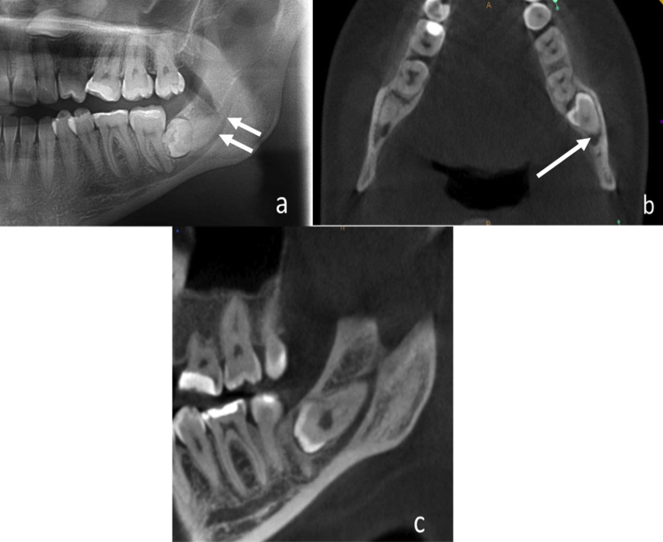
This study aimed to validate the accuracy of panoramic radiographic risk signs through detection of presence or absence of corticalization between an impacted mandibular third molar and the inferior alveolar canal on cone beam computed tomography (CBCT).
This retrospective study analyzed 210 impacted mandibular third molars from 135 patients (aged 17-51 years) who showed one or more of the seven previously established panoramic radiographic risk signs of inferior alveolar nerve exposure. These patients were referred for CBCT examination. Three-dimensional images were used to assess the canal position relative to the third molar, the proximity between the canal and third molar, and third molar angulation. The correlation of panoramic findings and CBCT was evaluated using a Chi-square test.
Panoramic findings of interruption of inferior alveolar canal wall, isolated or combined with one of these signs (darkening of third molar roots, narrowing of canal, and diversion of canal); darkening of the roots; and narrowing of canal were significantly correlated with direct contact between the inferior alveolar canal and impacted third molars on CBCT (P < 0.001).
Preoperative CBCT is recommended for cases showing interruption of canal wall; darkening of the roots or narrowing of the canal; or association between interruption and narrowing, diversion, or darkening of roots in a panoramic view. This study evaluated the risk relationship between the inferior alveolar nerve and impacted mandibular third molars, with the aim of reducing the occurrence of postoperative injury to the inferior alveolar nerve.
本研究旨在通过锥形束计算机断层扫描(CBCT)检测下颌阻生第三磨牙与下牙槽神经管之间是否存在骨皮质化,以验证全景X线片风险征象的准确性。
本回顾性研究分析了135例患者(年龄17 - 51岁)的210颗下颌阻生第三磨牙,这些患者表现出先前确定的下牙槽神经暴露的七个全景X线片风险征象中的一个或多个。这些患者被转诊进行CBCT检查。使用三维图像评估神经管相对于第三磨牙的位置、神经管与第三磨牙之间的距离以及第三磨牙的角度。使用卡方检验评估全景X线片结果与CBCT之间的相关性。
全景X线片显示下牙槽神经管壁中断,单独或与这些征象之一(第三磨牙牙根变黑、神经管变窄和神经管移位)合并;牙根变黑;以及神经管变窄,与CBCT显示的下牙槽神经管与阻生第三磨牙直接接触显著相关(P < 0.001)。
对于全景X线片显示神经管壁中断;牙根变黑或神经管变窄;或神经管壁中断与牙根变黑、变窄或移位相关的病例,建议术前进行CBCT检查。本研究评估了下牙槽神经与下颌阻生第三磨牙之间的风险关系,旨在减少术后下牙槽神经损伤的发生。