Pan Hong-Ming, Lang Wei-Ya, Yao Li-Jie, Wang Yan, Li Xiao-Ling
Department of Biochemistry, Qiqihar Medical University, Qiqihar 161000, Heilongjiang Province, China.
Department of Histology and Embryology, Qiqihar Medical University, Qiqihar 161000, Heilongjiang Province, China.
World J Gastrointest Oncol. 2019 Aug 15;11(8):622-633. doi: 10.4251/wjgo.v11.i8.622.
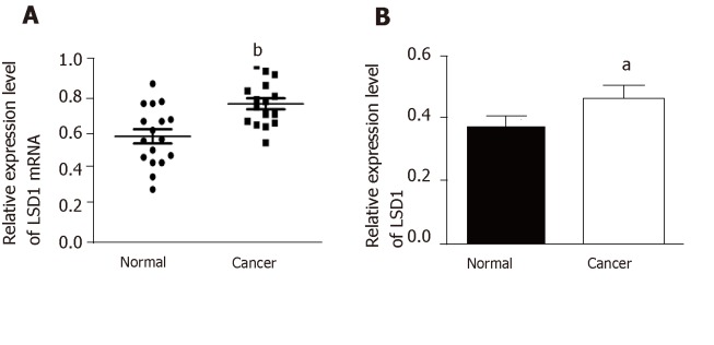
Histone Lysine Specific Demethylase 1 (LSD1) is the first histone demethylase to be discovered, which regulates various biological functions by making lysine of histone H3K4, H3K9 and non-histone substrates demethylated. Abnormal regulation of LSD1 is closely related to the occurrence and development of gastric cancer. The change of LSD1 expression level plays an important role in the proliferation and metastasis of gastric cancer cells. The study of its function and mechanism may provide a theoretical basis for early diagnosis and targeted therapy of gastric cancer.
To investigate the effect of downregulation of lysine-specific demethylase 1 (LSD1) expression on proliferation and invasion of gastric cancer cells and the possible regulatory mechanisms of the VEGF-C/PI3K/AKT signaling pathway.
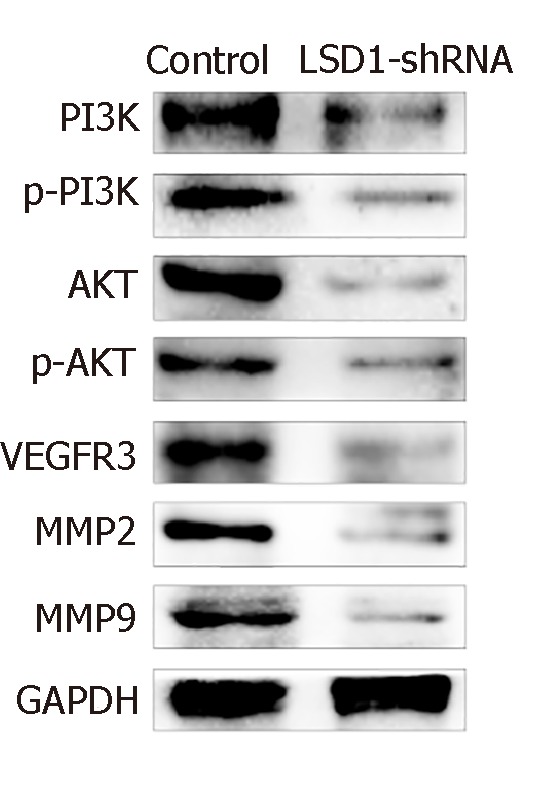
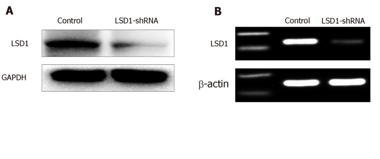
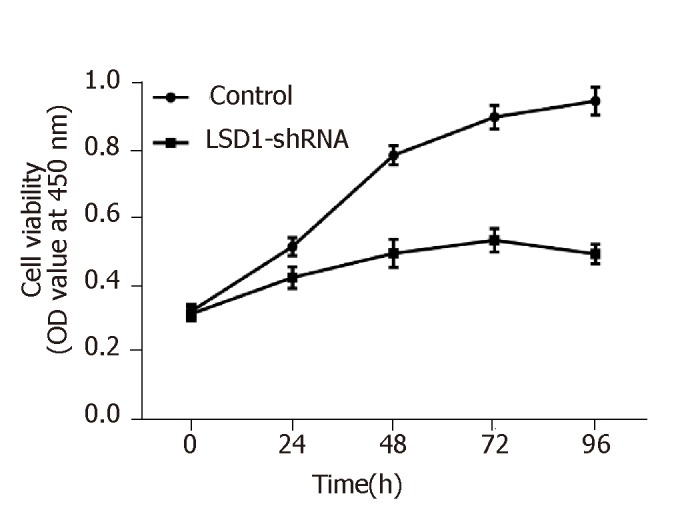
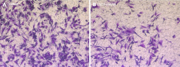
The LSD1-specific short hairpin RNA (shRNA) interference plasmid was transiently transfected, and expression of LSD1 was downregulated. The cell proliferation ability of LSD1 was observed by CCK-8 assay after downregulating expression of LSD1. Transwell invasion assay was used to observe the change of cell invasion ability after downregulating expression of LSD1. Expression of phosphorylated phosphoinositide 3-kinase (p-PI3K), PI3K, p-AKT, AKT, vascular endothelial growth factor receptor (VEGFR)-3, matrix metalloproteinase (MMP)-2 and MMP-9 in each group was detected by Western blotting.
The cell proliferation ability of transiently transfected LSD1-shRNA interference plasmid group was significantly lower than that of the control group ( < 0.05). Transwell invasion assay showed that the number of cells across the membrane of the LSD1-shRNA transfection group (238.451 ± 5.216) was significantly lower than that of the control group (49.268 ± 6.984) ( < 0.01). Western blotting showed that expression level of VEGF-C, p-PI3K, PI3K, p-AKT, AKT, VEGFR-3, MMP-2 and MMP-9 in the LSD1-shRNA group was significantly lower than that in the control group ( < 0.05).
Downregulation of LSD1 expression inhibits metastatic potential of gastric cancer cells, and VEGF-C-mediated activation of PI3K/AKT signaling pathway, which may be an important mechanism for inhibiting lymph node metastasis in gastric cancer cells.
组蛋白赖氨酸特异性去甲基化酶1(LSD1)是首个被发现的组蛋白去甲基化酶,通过使组蛋白H3K4、H3K9的赖氨酸及非组蛋白底物去甲基化来调节多种生物学功能。LSD1的异常调控与胃癌的发生发展密切相关。LSD1表达水平的变化在胃癌细胞的增殖和转移中起重要作用。对其功能及机制的研究可能为胃癌的早期诊断和靶向治疗提供理论依据。
探讨下调赖氨酸特异性去甲基化酶1(LSD1)表达对胃癌细胞增殖和侵袭的影响以及VEGF-C/PI3K/AKT信号通路可能的调控机制。
瞬时转染LSD1特异性短发夹RNA(shRNA)干扰质粒,下调LSD1表达。下调LSD1表达后,采用CCK-8法观察LSD1对细胞增殖能力的影响。采用Transwell侵袭实验观察下调LSD1表达后细胞侵袭能力的变化。通过蛋白质免疫印迹法检测各组中磷酸化磷脂酰肌醇3激酶(p-PI3K)、PI3K、p-AKT、AKT、血管内皮生长因子受体(VEGFR)-3、基质金属蛋白酶(MMP)-2和MMP-9的表达。
瞬时转染LSD1-shRNA干扰质粒组的细胞增殖能力显著低于对照组(<0.05)。Transwell侵袭实验显示,LSD1-shRNA转染组穿过膜的细胞数(238.451±5.216)显著低于对照组(49.268±6.984)(<0.01)。蛋白质免疫印迹法显示,LSD1-shRNA组中VEGF-C、p-PI3K、PI3K、p-AKT、AKT、VEGFR-3、MMP-2和MMP-9的表达水平显著低于对照组(<0.05)。
下调LSD1表达可抑制胃癌细胞的转移潜能,VEGF-C介导的PI3K/AKT信号通路激活可能是抑制胃癌细胞淋巴结转移的重要机制。