Department of Biomedical Laboratory Science, Faculty of Natural Sciences, NTNU-Norwegian University of Science and Technology, Trondheim, Norway.
Centre of Molecular Inflammation Research, Department of Clinical and Molecular Medicine, NTNU-Norwegian University of Science and Technology, Trondheim, Norway.
J Cachexia Sarcopenia Muscle. 2020 Feb;11(1):195-207. doi: 10.1002/jcsm.12489. Epub 2019 Aug 21.
The majority of patients with advanced cancer develop cachexia, a weight loss syndrome that severely reduces quality of life and limits survival. Our understanding of the underlying mechanisms that cause the condition is limited, and there are currently no treatment options that can completely reverse cachexia. Several tumour-derived factors and inflammatory mediators have been suggested to contribute to weight loss in cachectic patients. However, inconsistencies between studies are recurrent. Activin A and interleukin 6 (IL-6) are among the best studied factors that seem to be important, and several studies support their individual role in cachexia development.
We investigated the interplay between activin A and IL-6 in the cachexia-inducing TOV21G cell line, both in culture and in tumours in mice. We previously found that the human TOV21G cells secrete IL-6 that induces autophagy in reporter cells and cachexia in mice. Using this established cachexia cell model, we targeted autocrine activin A by genetic, chemical, and biological approaches. The secretion of IL-6 from the cancer cells was determined in both culture and tumour-bearing mice by a species-specific ELISA. Autophagy reporter cells were used to monitor the culture medium for autophagy-inducing activities, and muscle mass changes were evaluated in tumour-bearing mice.
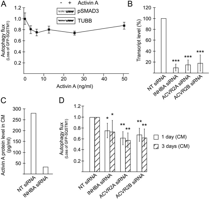
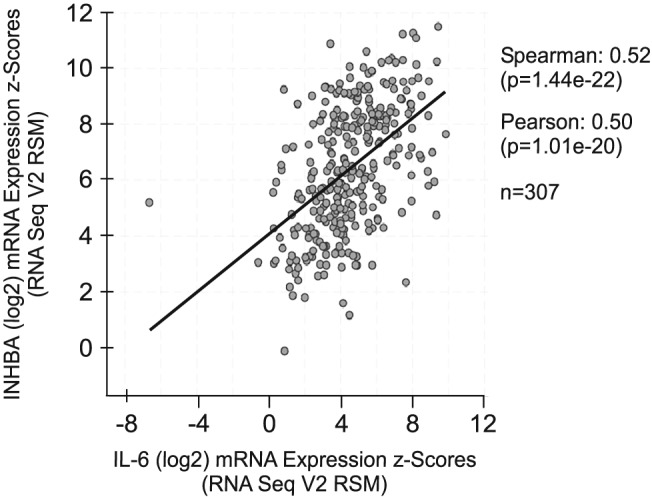
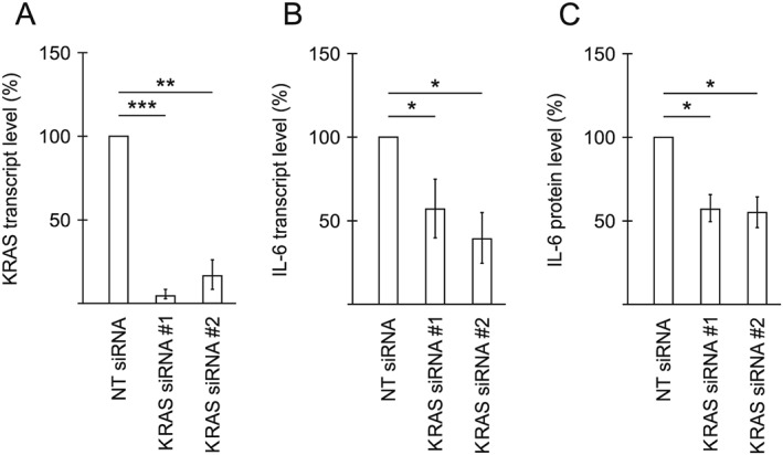
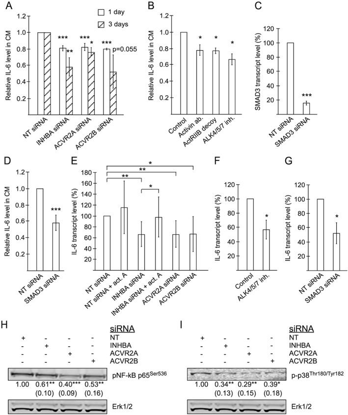
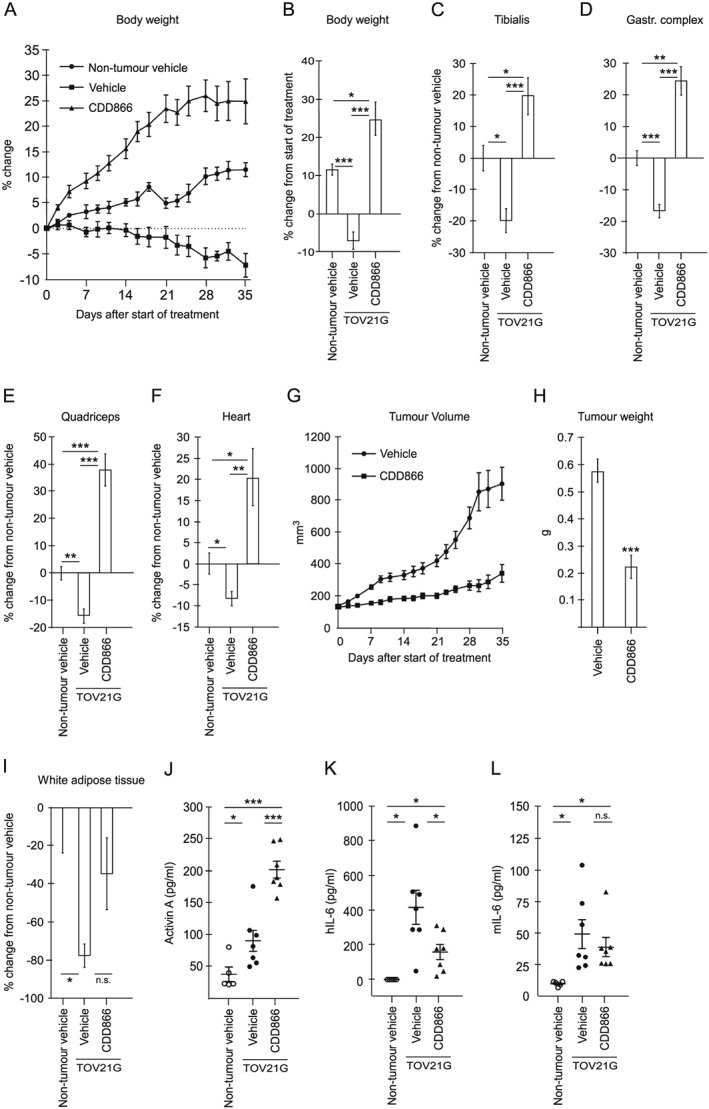
We show that activin A acts in an autocrine manner to promote the synthesis and secretion of IL-6 from cancer cells. By inhibiting activin A signalling, the production of IL-6 from the cancer cells is reduced by 40-50% (up to 42% reduction on protein level, P = 0.0048, and 48% reduction on mRNA level, P = 0.0308). Significantly reduced IL-6 secretion (P < 0.05) from the cancer cells is consistently observed when using biological, chemical, and genetic approaches to interfere with the autocrine activin A loop. Inhibiting activin signalling also reduces the ability of the cancer cells to accelerate autophagy in non-cancerous cells (up to 43% reduced autophagy flux, P = 0.0006). Coherent to the in vitro data, the use of an anti-activin receptor 2 antibody in cachectic tumour-bearing mice reduces serum levels of cancer cell-derived IL-6 by 62% (from 417 to 159 pg/mL, P = 0.03), and, importantly, it reverses cachexia and counteracts loss of all measured muscle groups (P < 0.0005).
Our data support a functional link between activin A and IL-6 signalling pathways and indicate that interference with activin A-induced IL-6 secretion from the tumour has therapeutic potential for cancer-induced cachexia.
大多数晚期癌症患者都会出现恶病质,这是一种严重降低生活质量并限制生存时间的体重减轻综合征。我们对导致这种疾病的潜在机制的了解是有限的,目前还没有可以完全逆转恶病质的治疗方法。有几种肿瘤衍生的因子和炎症介质被认为与恶病质患者的体重减轻有关。然而,研究之间的不一致性是反复出现的。激活素 A 和白细胞介素 6(IL-6)是研究最多的因子之一,它们似乎很重要,并且有几项研究支持它们在恶病质发展中的单独作用。
我们研究了激活素 A 和 IL-6 在诱导恶病质的 TOV21G 细胞系中的相互作用,包括在培养物中和在小鼠肿瘤中。我们之前发现,人类 TOV21G 细胞分泌的 IL-6 可诱导报告细胞自噬,并可诱导小鼠恶病质。使用这种已建立的恶病质细胞模型,我们通过遗传、化学和生物学方法靶向自分泌激活素 A。通过物种特异性 ELISA 测定培养物和荷瘤小鼠中癌细胞分泌的 IL-6。使用自噬报告细胞监测培养物中诱导自噬的活性,并评估荷瘤小鼠的肌肉质量变化。
我们表明,激活素 A 以自分泌方式促进癌细胞合成和分泌 IL-6。通过抑制激活素 A 信号,癌细胞产生的 IL-6 减少 40-50%(蛋白水平减少 42%,P=0.0048,mRNA 水平减少 48%,P=0.0308)。当使用生物、化学和遗传方法干扰自分泌激活素 A 环时,始终观察到癌细胞分泌的 IL-6 显著减少(P<0.05)。抑制激活素信号还降低了癌细胞加速非癌细胞自噬的能力(自噬通量减少 43%,P=0.0006)。与体外数据一致的是,在恶病质荷瘤小鼠中使用抗激活素受体 2 抗体可使癌细胞来源的 IL-6 血清水平降低 62%(从 417 降至 159 pg/mL,P=0.03),重要的是,它可逆转恶病质并抵消所有测量的肌肉群的损失(P<0.0005)。
我们的数据支持激活素 A 和 IL-6 信号通路之间的功能联系,并表明干扰肿瘤中激活素 A 诱导的 IL-6 分泌对癌症引起的恶病质具有治疗潜力。