Samreen Naziya, Lee Christine, Bhatt Asha, Carter Jodi, Hieken Tina, Adler Kalie, Zingula Shannon, Glazebrook Katrina N
Department of Radiology, Mayo Clinic Rochester, MN USA.
Department of Radiology, Laboratory Medicine and Pathology, Mayo Clinic Rochester, MN USA.
J Clin Imaging Sci. 2019 Apr 30;9:11. doi: 10.25259/JCIS_97_18. eCollection 2019.
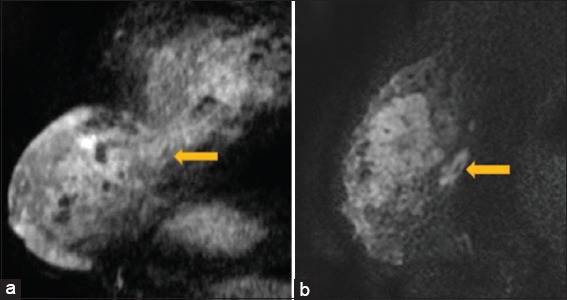
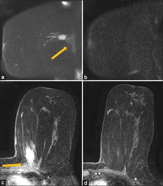
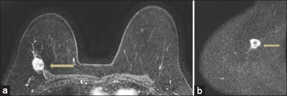
The purpose of this study is to evaluate diffusion weighted magnetic rsonance imaging (MRI) acquisitions in delineating posterior extent of breast tumors and in predicting chest wall invasion prior to treatment. To our knowledge, there has not been any literature specifically evaluating the utility of diffusion-weighted acquisitions in chest wall invasion of breast tumors.
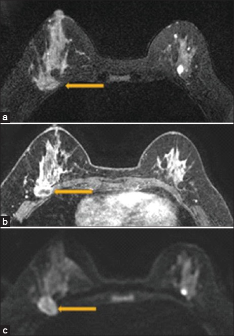
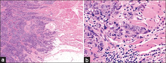
A retrospective review of our breast imaging database for keywords "chest wall invasion" and "breast MRI" was performed over the last 14 years. Diffusion sequences, T1 sequences (pre and post contrast), and T2 sequences were evaluated. Apparent diffusion coefficient (ADC) values in tumor and chest wall were assessed. Imaging findings were correlated with surgical pathology.
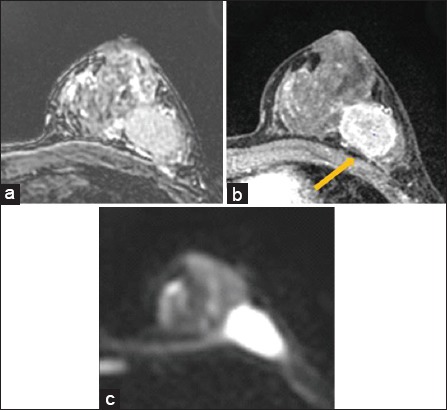
23 patients met inclusion criteria. All 23 had loss of fat plane on T2 sequences. 22/23 had loss of fat plane on postcontrast T1 sequences. Pectoralis muscle enhancement was present in 19/23 (83%) tumors and chest wall enhancement was present 9/23 (39%) tumors. Qualitative restricted diffusion within the pectoralis muscle was present in 18/23 (71%) tumors and in the chest wall was present in 8/23 (35%) tumors. Mean ADC values were 1.15 s/mm in the tumor and 1.29 s/mm in the chest wall. Sensitivity, specificity, positive predictive value and negative predictive value were 100%, 36%, 63%, and 100% for chest wall enhancement respectively and 69%, 36%, 61%, and 80% for chest wall diffusion-weighted imaging restriction respectively.
Diffusion weighted sequences can be helpful in characterizing chest wall invasion of breast tumors.
本研究的目的是评估扩散加权磁共振成像(MRI)在描绘乳腺肿瘤的后方范围以及在治疗前预测胸壁侵犯方面的应用。据我们所知,尚无文献专门评估扩散加权成像在乳腺肿瘤胸壁侵犯中的效用。
对我们的乳腺影像数据库进行回顾性研究,在过去14年中搜索关键词“胸壁侵犯”和“乳腺MRI”。评估扩散序列、T1序列(增强前后)和T2序列。评估肿瘤和胸壁中的表观扩散系数(ADC)值。将影像表现与手术病理结果进行关联。
23例患者符合纳入标准。所有23例在T2序列上均有脂肪平面消失。23例中有22例在增强后T1序列上有脂肪平面消失。23例中有19例(83%)肿瘤出现胸大肌强化,23例中有9例(39%)肿瘤出现胸壁强化。23例中有18例(71%)肿瘤在胸大肌内出现定性的扩散受限,23例中有8例(35%)肿瘤在胸壁出现扩散受限。肿瘤的平均ADC值为1.15 s/mm²,胸壁的平均ADC值为1.29 s/mm²。胸壁强化的敏感性、特异性、阳性预测值和阴性预测值分别为100%、36%、63%和100%,胸壁扩散加权成像受限的敏感性、特异性、阳性预测值和阴性预测值分别为69%、36%、61%和80%。
扩散加权序列有助于对乳腺肿瘤的胸壁侵犯进行特征性描述。