Afify Said M, Hassan Ghmkin, Osman Amira, Calle Anna Sanchez, Nawara Hend M, Zahra Maram Hussein, El-Ghlban Samah, Mansour Hager, Alam Md Jahangir, Abu Quora Hagar A, Du Juan, Seno Akimasa, Iwasaki Yoshiaki, Seno Masaharu
Department of Medical Bioengineering, Graduate School of Natural Science and Technology, Okayama University, Okayama 700-8530, Japan.
Division of Biochemistry, Faculty of Science, Menoufia University, Shebin El Koum, Menoufia 32511, Egypt.
Bioengineering (Basel). 2019 Aug 23;6(3):73. doi: 10.3390/bioengineering6030073.
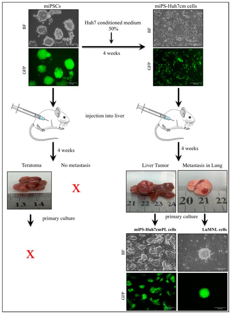
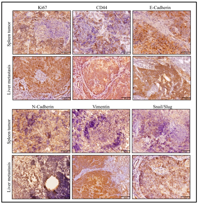
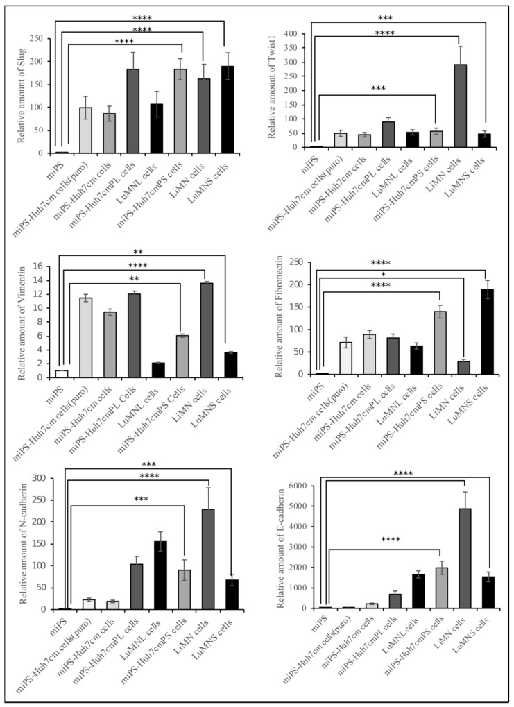
Metastasis develops when cancer cells spread from the primary site of a malignant tumor to the surrounding and distant tissues, and it is the most critical problem in cancer treatment. Our group developed cancer stem cells (CSCs) from induced pluripotent stem cells (iPSCs) in the presence of a conditioned medium (CM) of cancer-derived cells. The CSCs were characterized by the formation of malignant tumors in vivo, followed by metastasis. In this study, CSCs converted from mouse iPSCs in the presence of CM from hepatocellular carcinoma (HCC) cell line Huh7 cells. These converted cells (miPS-Huh7cm cells) were established as the metastatic cells. The generated CSCs were injected into the liver or spleen of nude mice. Almost one month after transplantation, the tumors were excised, and the primary cultured cells derived from the malignant tumors and metastatic nodules were evaluated by stemness and metastatic markers to compare their differences. The miPS-Huh7cm cells exhibited metastatic potential, and efficiently formed malignant tumors with lung and/or liver lesions in vivo, whereas the injected miPS formed teratoma. The primary cultured cells derived from the malignant tumors and metastatic nodules sustained the expression of stemness markers, such as Nanog, Klf4 and c-Myc, and acquired cancer stem markers, such as CD90, CD44 and ALDH1. Simultaneously, the expression of metastatic markers, such as Slug, Twist1 and vimentin, in primary cells derived from the malignant tumors, was higher than in metastatic nodules. The CSCs derived from iPSCs, forming malignant tumors and displaying high metastasis, will provide a good animal model to study the mechanisms of metastasis.
当癌细胞从恶性肿瘤的原发部位扩散到周围及远处组织时,就会发生转移,这是癌症治疗中最关键的问题。我们的研究小组在癌症来源细胞的条件培养基(CM)存在的情况下,从诱导多能干细胞(iPSC)中培养出了癌症干细胞(CSC)。这些CSC的特征是在体内形成恶性肿瘤,随后发生转移。在本研究中,在来自肝癌(HCC)细胞系Huh7细胞的CM存在下,从小鼠iPSC转化得到了CSC。这些转化细胞(miPS-Huh7cm细胞)被确立为转移细胞。将产生的CSC注射到裸鼠的肝脏或脾脏中。移植后近一个月,切除肿瘤,并通过干性和转移标志物对源自恶性肿瘤和转移结节的原代培养细胞进行评估,以比较它们的差异。miPS-Huh7cm细胞具有转移潜能,在体内能有效地形成伴有肺和/或肝损伤的恶性肿瘤,而注射的miPS则形成畸胎瘤。源自恶性肿瘤和转移结节的原代培养细胞持续表达干性标志物,如Nanog、Klf4和c-Myc,并获得癌症干细胞标志物,如CD90、CD44和ALDH1。同时,源自恶性肿瘤的原代细胞中转移标志物,如Slug、Twist1和波形蛋白的表达高于转移结节中的表达。源自iPSC的CSC形成恶性肿瘤并表现出高转移特性,将为研究转移机制提供良好的动物模型。