Huan Hong-Bo, Yang Da-Peng, Wen Xu-Dong, Chen Xue-Jiao, Zhang Liang, Wu Li-Li, Bie Ping, Xia Feng
Institute of Hepatobiliary Surgery, Southwest Cancer Center, Southwest Hospital, Third Military Medical University, Chongqing, 400038, China.
Laboratory of Biotherapy of Cancer, Southwest Cancer Center, Southwest Hospital, Third Military Medical University, Chongqing, 400038, China.
J Exp Clin Cancer Res. 2017 Jun 24;36(1):86. doi: 10.1186/s13046-017-0559-4.
Homeobox B7 (HOXB7) has been identified associated with poor prognosis of hepatocellular carcinoma (HCC). However, the specific mechanism by which HOXB7 promotes the malignant progression of HCC remains to be determined.
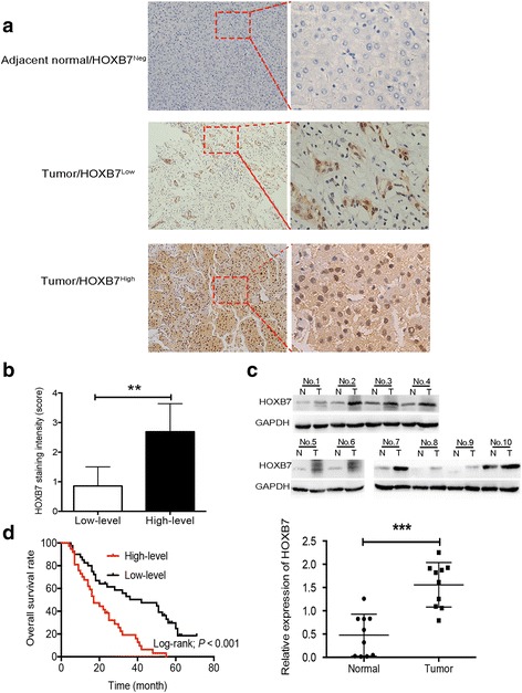
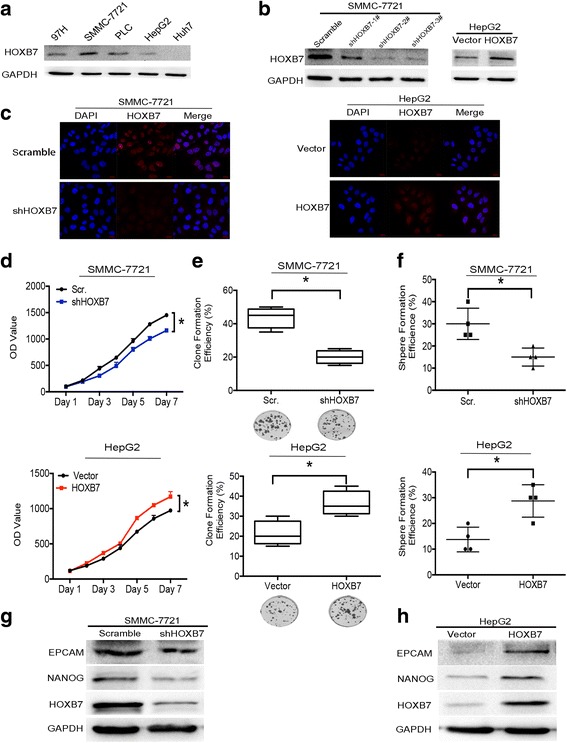
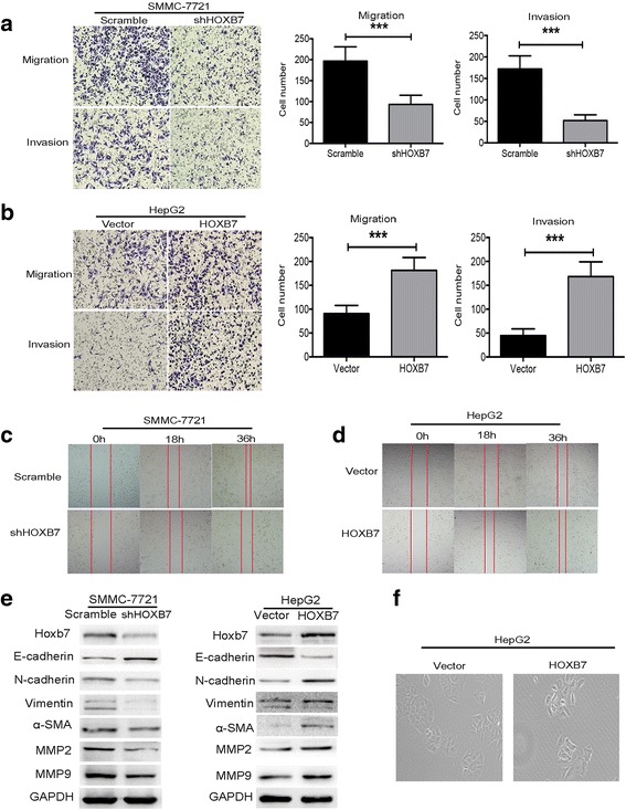
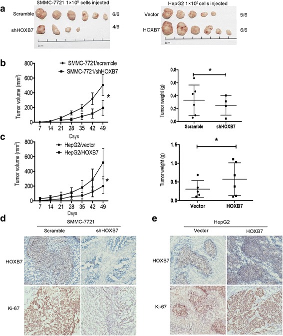
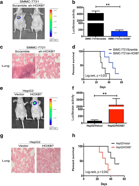
Immunohistochemistry (IHC) was used to detect the expression level of HOXB7 in 77-paired HCC tissue samples, and the correlation between HOXB7 and HCC prognosis was assessed. The location of HOXB7 was confirmed by immunofluorescence. Cell Titer-Blue assay was used to assess the proliferation of hepatoma cells. The stem-like properties of hepatoma cells were analysed by sphere formation and clone formation assays. The effect of HOXB7 on expression of cancer stem cell markers was evaluated. Transwell and wound-healing assays were performed to estimate the invasion and migration abilities of hepatoma cells. A xenograft tumor model was established in nude mice to assess the role of HOXB7 in tumor growth. Bioluminescence imaging was used to survey the effect of HOXB7 on the metastatic ability of hepatoma cells in vivo.
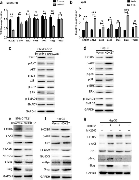
Higher expression of HOXB7 was detected in HCC tissues compared with noncancerous tissues and significantly associated with poor prognosis of HCC. In addition, HOXB7 knockdown suppressed the cell proliferation, clone formation, sphere formation, invasion and migration of hepatoma cells in vitro; conversely, these biological abilities of hepatoma cells were enhanced by HOXB7 overexpression. Moreover, the cancer stem cell markers EPCAM and NANOG were up-regulated by HOXB7. The role of HOXB7 in promoting tumor growth and metastasis was verified in vivo. Further investigation revealed that c-Myc and Slug expression was elevated by HOXB7 and the AKT pathway was activated.
Overexpression of HOXB7 was significantly correlated with poor prognosis of HCC. HOXB7 up-regulated c-Myc and Slug expression via the AKT pathway to promote the acquisition of stem-like properties and facilitate epithelial-mesenchymal transition of hepatoma cells, accelerating the malignant progression of HCC.
同源盒B7(HOXB7)已被证实与肝细胞癌(HCC)的不良预后相关。然而,HOXB7促进HCC恶性进展的具体机制仍有待确定。
采用免疫组织化学(IHC)检测77对HCC组织样本中HOXB7的表达水平,并评估HOXB7与HCC预后的相关性。通过免疫荧光确定HOXB7的定位。采用细胞活力检测法评估肝癌细胞的增殖情况。通过成球和克隆形成试验分析肝癌细胞的干性特征。评估HOXB7对癌症干细胞标志物表达的影响。进行Transwell和伤口愈合试验以评估肝癌细胞的侵袭和迁移能力。在裸鼠中建立异种移植肿瘤模型,以评估HOXB7在肿瘤生长中的作用。利用生物发光成像技术在体内观察HOXB7对肝癌细胞转移能力的影响。
与癌旁组织相比,HCC组织中HOXB7表达更高,且与HCC的不良预后显著相关。此外,HOXB7基因敲低抑制了体外肝癌细胞的增殖、克隆形成、成球、侵袭和迁移;相反,HOXB7过表达增强了肝癌细胞的这些生物学能力。此外,HOXB7上调了癌症干细胞标志物EPCAM和NANOG的表达。HOXB7在促进肿瘤生长和转移方面的作用在体内得到了验证。进一步研究发现,HOXB7升高了c-Myc和Slug的表达,并激活了AKT信号通路。
HOXB7过表达与HCC的不良预后显著相关。HOXB7通过AKT信号通路上调c-Myc和Slug的表达,促进肝癌细胞获得干性特征并促进上皮-间质转化,加速HCC的恶性进展。