Cardenas Daviel, Bhalchandra Seema, Lamisere Hymlaire, Chen Ying, Zeng Xi-Lei, Ramani Sasirekha, Karandikar Umesh C, Kaplan David L, Estes Mary K, Ward Honorine D
Tufts Medical Center, Boston, MA, USA.
Tufts University Sackler School of Graduate Biomedical Sciences, Boston, MA, USA.
Methods Mol Biol. 2020;2052:373-402. doi: 10.1007/978-1-4939-9748-0_21.
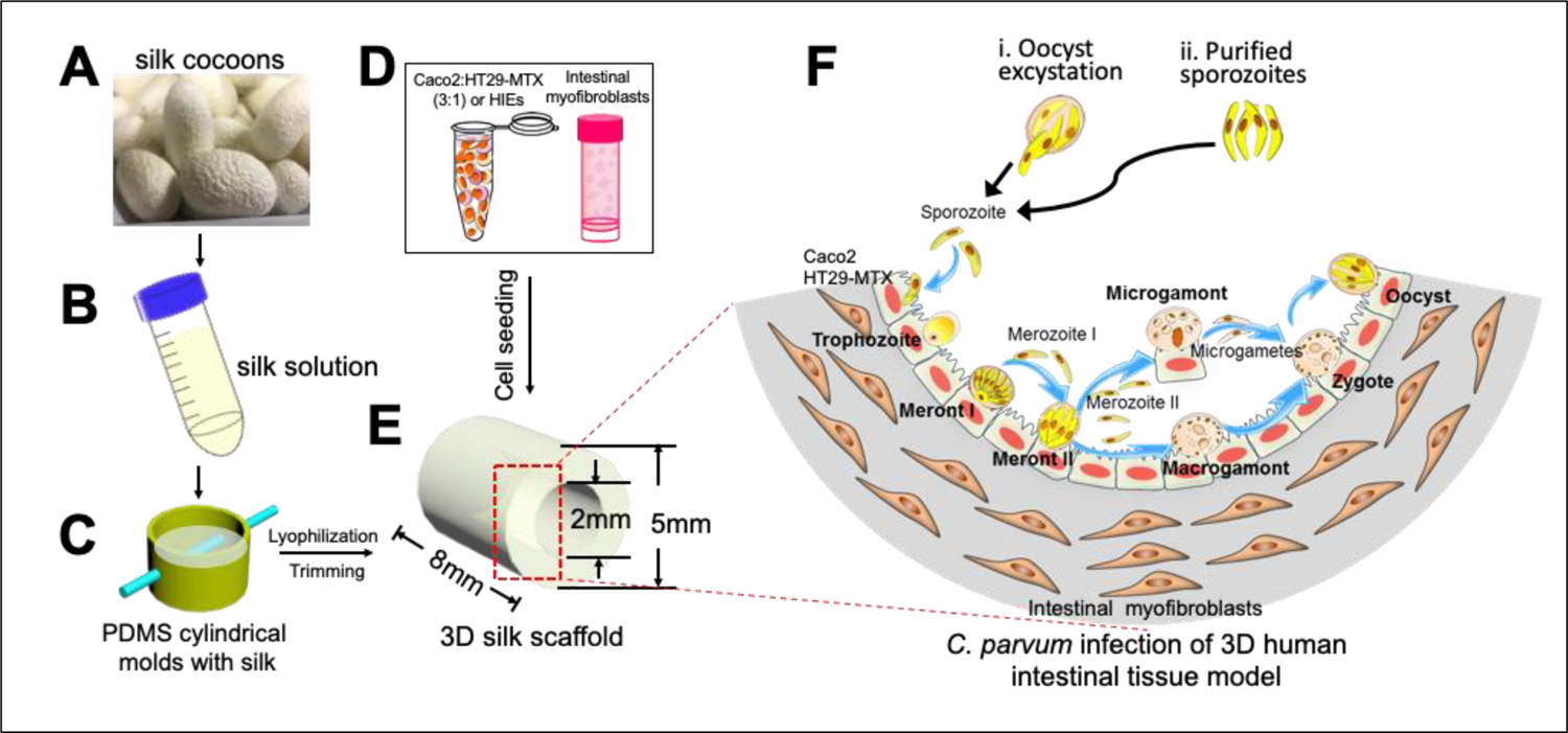
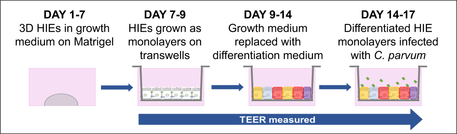
Conventional cell cultures utilizing transformed or immortalized cell lines or primary human epithelial cells have played a fundamental role in furthering our understanding of Cryptosporidium infection. However, they remain inadequate with respect to their inability to emulate in vivo conditions, support long-term growth, and complete the life cycle of the parasite. Previously, we developed a 3D silk scaffold-based model using transformed human intestinal epithelial cells (IECs). This model supported C. parvum infection for up to 2 weeks and resulted in completion of the life cycle of the parasite. However, transformed IECs are not representative of primary human IEC.Human intestinal enteroids (HIEs) are cultures derived from crypts that contain Lgr5 stem cells isolated from human biopsies or surgical intestinal tissues; these established multicellular cultures can be induced to differentiate into enterocytes, enteroendocrine cells, goblet cells, Paneth cells, and tuft cells. HIEs better represent human intestinal structure and function than immortalized IEC lines. Recently, significant progress has been made in the development of technologies to culture HIEs in vitro. When grown in a 3D matrix, HIEs provide a spatial organization resembling the native human intestinal epithelium. Additionally, they can be dissociated and grown as monolayers in tissue culture plates, permeable supports or silk scaffolds that enable mechanistic studies of pathogen infections. They can also be co-cultured with other human cells such as macrophages and myofibroblasts. The HIEs grown in these novel culture systems recapitulate the physiology, the 3D architecture, and functional diversity of native intestinal epithelium and provide a powerful and promising new tool to study Cryptosporidium-host cell interactions and screen for interventions ex vivo. In this chapter, we describe the 3D silk scaffold-based model using transformed IEC co-cultured with human intestinal myofibroblasts and 2D and 3D HIE-derived models of Cryptosporidium, also co-cultured with human intestinal myofibroblasts.
利用转化或永生化细胞系或原代人上皮细胞的传统细胞培养,在增进我们对隐孢子虫感染的理解方面发挥了重要作用。然而,它们在模拟体内条件、支持长期生长以及完成寄生虫生命周期方面仍存在不足。此前,我们使用转化的人肠上皮细胞(IECs)开发了一种基于3D丝支架的模型。该模型支持微小隐孢子虫感染长达2周,并导致寄生虫生命周期的完成。然而,转化的IECs并不代表原代人IEC。人肠道类器官(HIEs)是从含有从人活检组织或手术肠道组织中分离出的Lgr5干细胞的隐窝中培养而来的;这些已建立的多细胞培养物可被诱导分化为肠上皮细胞、肠内分泌细胞、杯状细胞、潘氏细胞和簇状细胞。与永生化IEC系相比,HIEs能更好地代表人类肠道结构和功能。最近,体外培养HIEs的技术取得了重大进展。当在3D基质中生长时,HIEs提供了一种类似于天然人类肠上皮的空间组织。此外,它们可以解离并在组织培养板、可渗透支持物或丝支架中作为单层生长,从而能够对病原体感染进行机制研究。它们还可以与其他人类细胞如巨噬细胞和成肌纤维细胞共培养。在这些新型培养系统中生长的HIEs概括了天然肠上皮的生理学、3D结构和功能多样性,并为研究隐孢子虫与宿主细胞相互作用以及在体外筛选干预措施提供了一个强大且有前景的新工具。在本章中,我们描述了使用与人类肠道成肌纤维细胞共培养的转化IEC构建的基于3D丝支架的模型,以及同样与人类肠道成肌纤维细胞共培养的隐孢子虫的2D和3D HIE衍生模型。