Integrated Program in Neuroscience, McGill University, Montreal, Quebec, Canada.
Computational Brain Anatomy Laboratory, Cerebral Imaging Centre, Douglas Mental Health University Institute, Verdun, Quebec, Canada.
Hum Brain Mapp. 2019 Dec 15;40(18):5269-5288. doi: 10.1002/hbm.24771. Epub 2019 Aug 26.
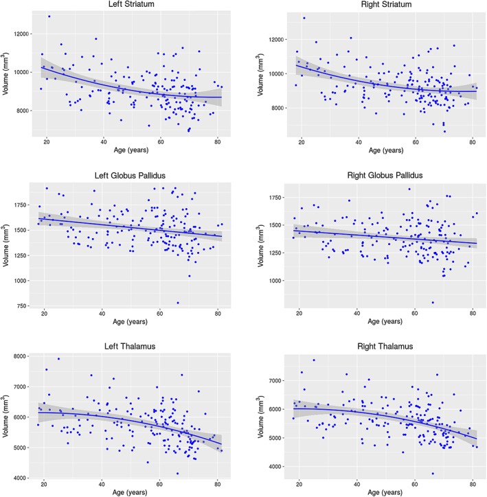
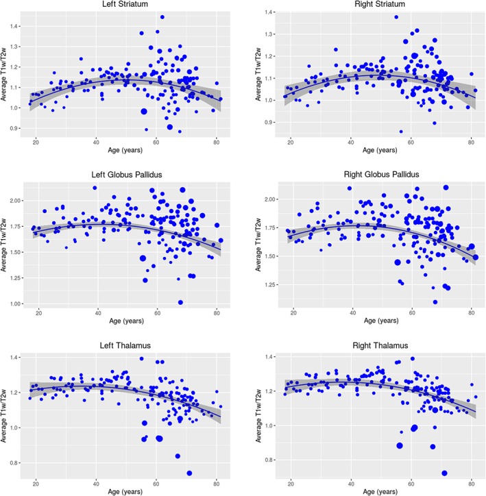
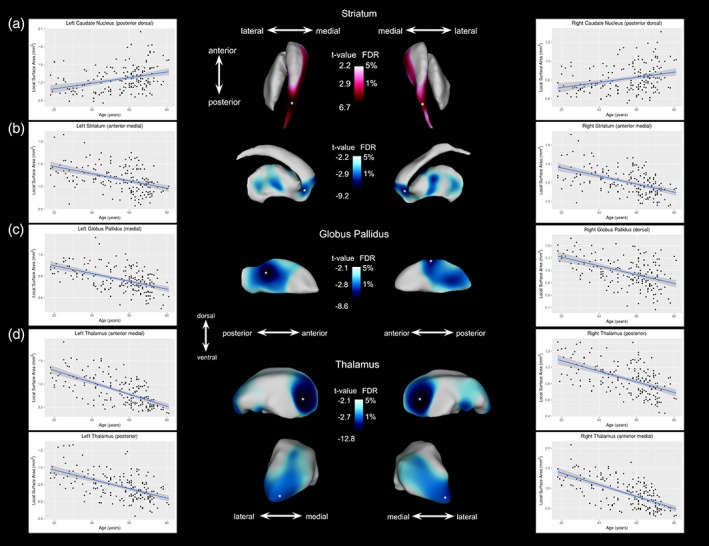
While numerous studies have used magnetic resonance imaging (MRI) to elucidate normative age-related trajectories in subcortical structures across the human lifespan, there exists substantial heterogeneity among different studies. Here, we investigated the normative relationships between age and morphology (i.e., volume and shape), and microstructure (using the T1-weighted/T2-weighted [T1w/T2w] signal ratio as a putative index of myelin and microstructure) of the striatum, globus pallidus, and thalamus across the adult lifespan using a dataset carefully quality controlled, yielding a final sample of 178 for the morphological analyses, and 162 for the T1w/T2w analyses from an initial dataset of 253 healthy subjects, aged 18-83. In accordance with previous cross-sectional studies of adults, we observed age-related volume decrease that followed a quadratic relationship between age and bilateral striatal and thalamic volumes, and a linear relationship in the globus pallidus. Our shape indices consistently demonstrated age-related posterior and medial areal contraction bilaterally across all three structures. Beyond morphology, we observed a quadratic inverted U-shaped relationship between T1w/T2w signal ratio and age, with a peak value occurring in middle age (at around 50 years old). After permutation testing, the Akaike information criterion determined age relationships remained significant for the bilateral globus pallidus and thalamus, for both the volumetric and T1w/T2w analyses. Our findings serve to strengthen and expand upon previous volumetric analyses by providing a normative baseline of morphology and microstructure of these structures to which future studies investigating patients with various disorders can be compared.
虽然许多研究已经使用磁共振成像(MRI)来阐明人类一生中皮质下结构的正常年龄相关轨迹,但不同研究之间存在很大的异质性。在这里,我们使用经过仔细质量控制的数据集,调查了纹状体、苍白球和丘脑的形态(即体积和形状)和微观结构(使用 T1 加权/T2 加权 [T1w/T2w] 信号比作为髓鞘和微观结构的假定指标)与成人寿命之间的正常关系,最终的形态分析样本为 178 个,T1w/T2w 分析样本为 162 个,来自 253 名健康受试者的初始数据集,年龄在 18-83 岁之间。与之前成年人的横断面研究一致,我们观察到与年龄相关的体积减少,这种减少遵循双侧纹状体和丘脑体积与年龄之间的二次关系,以及苍白球中的线性关系。我们的形状指数一致地显示出双侧所有三个结构的后内侧区域随年龄缩小。除了形态学,我们还观察到 T1w/T2w 信号比与年龄之间存在二次倒 U 形关系,峰值出现在中年(约 50 岁左右)。经过置换检验,Akaike 信息准则确定年龄关系对于双侧苍白球和丘脑仍然具有统计学意义,无论是在体积分析还是 T1w/T2w 分析中都是如此。我们的研究结果为之前的体积分析提供了支持和扩展,为这些结构的形态学和微观结构提供了一个正常的基线,未来研究各种疾病患者的研究可以与之进行比较。