Or Chris, Heier Jeffrey S, Boyer David, Brown David, Shah Sumit, Alibhai Agha Yasin, Fujimoto James G, Waheed Nadia
1New England Eye Center, Tufts Medical Center, 260 Tremont Street, Biewend Building, 9-11th Floor, Boston, MA 02111 USA.
2Ophthalmic Consultants of Boston, Boston, MA USA.
Int J Retina Vitreous. 2019 Aug 20;5:36. doi: 10.1186/s40942-019-0187-6. eCollection 2019.
To investigate whether neovascularization may arise and be detectable in drusen, as reported in histopathologic studies, by OCTA prior to developing exudation and to assess its prevalence in a cohort of patients with intermediate AMD.
Retrospective cross-sectional study of 128 patients with intermediate AMD recruited as part of a separate ongoing clinical trial conducted at multiple large tertiary referral retina clinics. One hundred and twenty-eight consecutive patients with exudative AMD in one eye and intermediate non-exudative AMD in the fellow eye were enrolled and analyzed between September 2015 and March 2017.
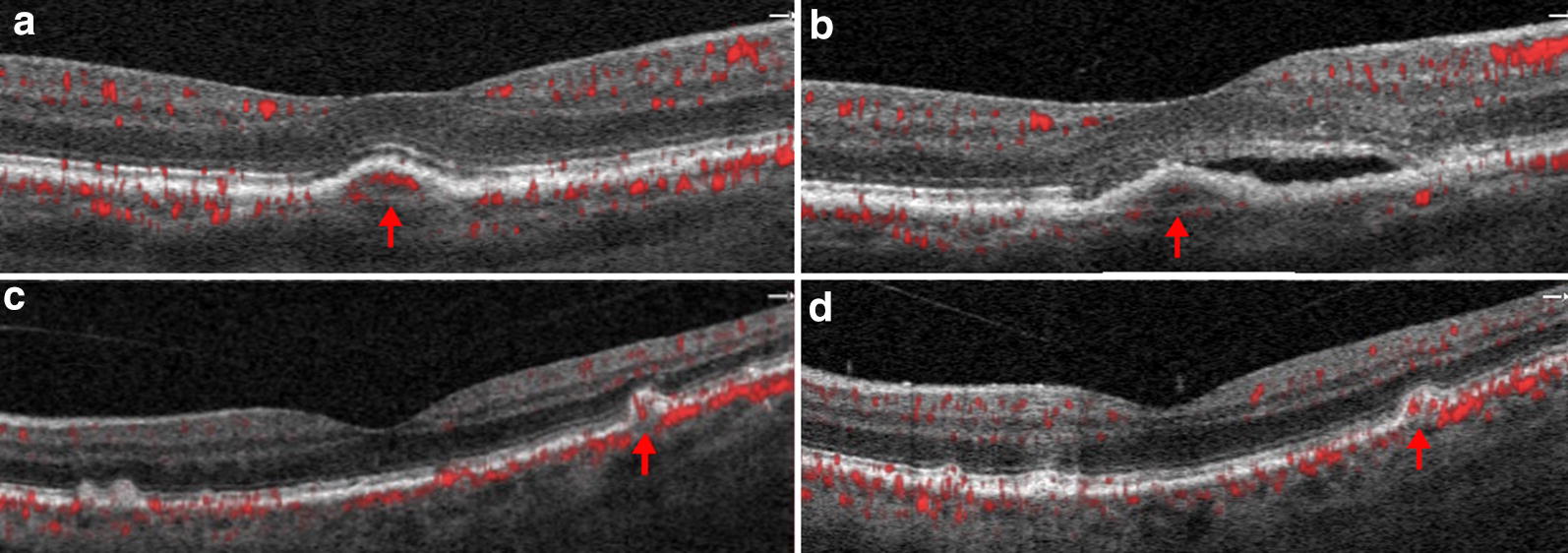
SD-OCTA identified vascularization within drusen in 7 of 128 eyes, for a prevalence of 5.5%. A total of 12 instances of vascularized drusen were noted. Out of the 12 vascularized drusen noted, 7 were located in the parafoveal region or subfoveal region and 5 was in the extrafoveal region. 9 of 12 instances of vascularized drusen exhibited a uniform sub-RPE hyperreflectivity, whilst 3 of 12 exhibited more heterogenous reflectivity. In all 12 instances, FA images failed to identify the neovascular nature of vascularized drusen.
Our results demonstrate the utility of SD-OCTA for the diagnosis of vascularized drusen in patients with intermediate non-exudative AMD. Longitudinal studies are needed to delineate the evolution and conversion risk of these lesions over time, which can be of substantial clinical relevance.
正如组织病理学研究所报道的,探讨在渗出发生之前,光学相干断层扫描血管造影(OCTA)是否能检测到玻璃膜疣中新生血管的出现,并评估其在一组中度年龄相关性黄斑变性(AMD)患者中的患病率。
对128例中度AMD患者进行回顾性横断面研究,这些患者是在多个大型三级转诊视网膜诊所进行的一项正在进行的独立临床试验中招募的。2015年9月至2017年3月期间,纳入并分析了128例一只眼为渗出性AMD且另一只眼为中度非渗出性AMD的连续患者。
光谱域光学相干断层扫描血管造影(SD-OCTA)在128只眼中的7只眼中检测到玻璃膜疣内有血管形成,患病率为5.5%。共记录到12例血管化玻璃膜疣。在记录到的12例血管化玻璃膜疣中,7例位于黄斑旁区域或黄斑下区域,5例位于黄斑外区域。12例血管化玻璃膜疣中有9例表现为均匀的视网膜色素上皮(RPE)下高反射,而12例中有3例表现出更多的异质性反射。在所有12例中,荧光素血管造影(FA)图像均未能识别出血管化玻璃膜疣的新生血管性质。
我们的结果证明了SD-OCTA在诊断中度非渗出性AMD患者血管化玻璃膜疣中的实用性。需要进行纵向研究来描述这些病变随时间的演变和转化风险,这可能具有重要的临床意义。