Iwasaki Motoyuki, Yokohama Takumi, Oura Daisuke, Furuya Shou, Niiya Yoshimasa, Okuaki Tomoyuki
Department of Neurosurgery, Otaru General Hospital, Otaru, Hokkaido, Japan.
Department of Radiology, Otaru General Hospital, Otaru, Hokkaido, Japan.
World Neurosurg X. 2019 Jul 26;4:100056. doi: 10.1016/j.wnsx.2019.100056. eCollection 2019 Oct.
Diffusion tensor imaging (DTI) is widely used; however, most of the prior studies have resulted in presurgical decreased fractional anisotropy (FA) values in patients with cervical spondylotic myelopathy (CSM). We used ZOOM DTI and could acquire highly accurate FA values during perioperative periods, which indicated different insights than preceding studies. The objective of this study was to assess the perioperative FA change in patients with CSM and determine the prognostic factor.
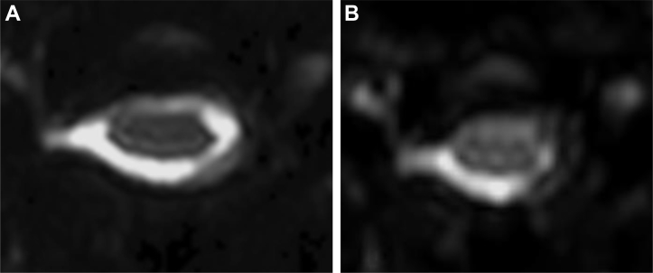
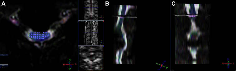
Twenty-eight patients with CSM and healthy control subjects were enrolled in this study. Twenty patients (71%) had intracordal high intensity before surgery. All patients underwent decompressive surgery. ZOOM DTI and the Japanese Orthopaedic Association (JOA) assessment were performed before and after surgery. The region of interest was manually contoured to omit the surrounding cerebrospinal fluid. The axial plane of the most stenotic cervical level was assessed.
FA values before surgery and at 1 week after surgery, and FA values at 1 week after surgery and at 6 months after surgery differed significantly as determined. The FA values of patients with intracordal high intensity significantly decreased after surgery and significantly increased from 1 week to 6 months, whereas those of patients without intracordal high intensity did not significantly change. JOA scores at 6 months after surgery (13.1) improved significantly compared with JOA scores before surgery (10.8). Only FA values at 1 week after surgery had a significant positive relationship with JOA scores presurgery and at 6 months after surgery.
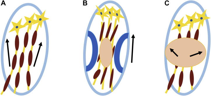
The presurgical FA value in patients with CSM did not differ from that of normal control subjects, but significantly decreased after surgery, and significantly increased 6 months after surgery. We concluded that the postsurgical FA value approximates the proper state of the damaged cord and the presurgical FA value includes a masked effect as an aligned fiber effect because of compression by degenerative construction. Only the FA value at 1 week had a significant positive relationship with the JOA score presugery and at 6 months, which established that the postsurgical FA value may be a more accurate prognostic factor than the presurgical FA value.
扩散张量成像(DTI)被广泛应用;然而,大多数先前的研究显示,脊髓型颈椎病(CSM)患者术前的分数 anisotropy(FA)值会降低。我们使用了 ZOOM DTI,能够在围手术期获得高度准确的 FA 值,这与之前的研究结果有所不同。本研究的目的是评估 CSM 患者围手术期 FA 的变化,并确定预后因素。
本研究纳入了 28 例 CSM 患者和健康对照者。20 例患者(71%)术前脊髓内有高强度信号。所有患者均接受了减压手术。在手术前后进行了 ZOOM DTI 和日本骨科协会(JOA)评估。手动勾勒感兴趣区域以排除周围的脑脊液。评估最狭窄颈椎节段的轴位平面。
术前与术后 1 周的 FA 值,以及术后 1 周与术后 6 个月的 FA 值差异显著。脊髓内有高强度信号的患者术后 FA 值显著降低,从术后 1 周到 6 个月显著升高,而脊髓内无高强度信号的患者 FA 值无显著变化。术后 6 个月的 JOA 评分(13.1)与术前 JOA 评分(10.8)相比有显著改善。仅术后 1 周的 FA 值与术前及术后 6 个月的 JOA 评分有显著正相关。
CSM 患者术前的 FA 值与正常对照者无差异,但术后显著降低,术后 6 个月显著升高。我们得出结论,术后 FA 值接近受损脊髓的正常状态,术前 FA 值因退变结构的压迫而存在作为排列纤维效应的掩盖效应。仅术后 1 周的 FA 值与术前及术后 6 个月的 JOA 评分有显著正相关,这表明术后 FA 值可能是比术前 FA 值更准确的预后因素。