Aging & Metabolism Research Program, Oklahoma Medical Research Foundation, Oklahoma City, OK, USA.
Advanced Magnetic Resonance Center, Oklahoma Medical Research Foundation, Oklahoma City, OK, USA.
Redox Biol. 2019 Sep;26:101308. doi: 10.1016/j.redox.2019.101308. Epub 2019 Aug 21.
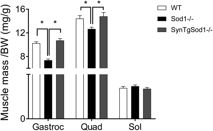
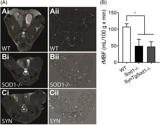
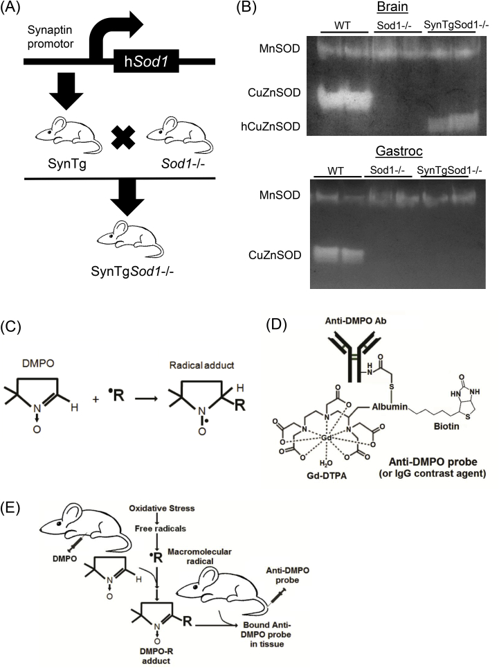
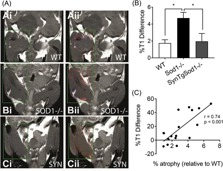
Mitochondrial dysfunction, reactive oxygen species (ROS) and oxidative damage have been implicated to play a causative role in age-related skeletal muscle atrophy and weakness (i.e. sarcopenia). Mice lacking the superoxide scavenger CuZnSOD (Sod1) exhibit high levels of oxygen-derived radicals and oxidative damage, associated with neuronal and muscular phenotypes consistent with sarcopenia. We used magnetic resonance imaging (MRI) technology combined with immunospin-trapping (IST) to measure in vivo free radical levels in skeletal muscle from wildtype, Sod1 and SynTgSod1 mice, a mouse model generated using targeted expression of the human Sod1 transgene specifically in neuronal tissues to determine the impact of motor neuron degeneration in muscle atrophy. By combining the spin trap DMPO (5,5-dimethyl-1-pyrroline N-oxide) and molecular MRI (mMRI), we monitored the level of free radicals in mouse hindlimb muscle. The level of membrane-bound macromolecular radicals in the quadriceps muscle was elevated by ~3-fold in Sod1 mice, but normalized to wildtype levels in SynTgSod1 rescue mice. Skeletal muscle mass was reduced by ~25-30% in Sod1 mice, but fully reversed in muscle from SynTgSod1 mice. Using perfusion MRI we also measured the dynamics of blood flow within mouse hindlimb. Relative muscle blood flow in Sod1 is decreased to ~50% of wildtype and remained low in the SynTgSod1 mice. Our findings are significant in that we have shown for the first time that in vivo free radical production in skeletal muscle is directly correlated to muscle atrophy in an experimental model of oxidative stress. Neuron-specific expression of CuZnSOD reverses the in vivo free radical production in skeletal muscle in the Sod1 mouse model and prevents muscle atrophy. These results further support the feasibility of using in vivo assessments of redox status in the progression of a pathological process such as sarcopenia. This approach can also be valuable for evaluating responses to pharmacologic interventions.
线粒体功能障碍、活性氧(ROS)和氧化损伤被认为在与年龄相关的骨骼肌萎缩和无力(即肌少症)中起因果作用。缺乏超氧化物清除剂 CuZnSOD(Sod1)的小鼠表现出高水平的氧衍生自由基和氧化损伤,与神经元和肌肉表型一致,符合肌少症的特征。我们使用磁共振成像(MRI)技术结合免疫自旋捕获(IST)来测量野生型、Sod1 和 SynTgSod1 小鼠骨骼肌中的体内自由基水平,SynTgSod1 小鼠是使用靶向表达人类 Sod1 转基因的方法在神经元组织中特异性生成的一种小鼠模型,以确定运动神经元退化对肌肉萎缩的影响。通过结合自旋捕获剂 DMPO(5,5-二甲基-1-吡咯啉 N-氧化物)和分子 MRI(mMRI),我们监测了小鼠后肢肌肉中的自由基水平。在 Sod1 小鼠中,四头肌中膜结合大分子自由基的水平升高了约 3 倍,但在 SynTgSod1 挽救小鼠中恢复到野生型水平。Sod1 小鼠的骨骼肌质量减少了约 25-30%,但在 SynTgSod1 小鼠的肌肉中完全逆转。使用灌注 MRI,我们还测量了小鼠后肢内的血流动力学。Sod1 中的相对肌肉血流减少到约野生型的 50%,而在 SynTgSod1 小鼠中仍然较低。我们的发现具有重要意义,因为我们首次表明,在氧化应激的实验模型中,骨骼肌中的体内自由基产生与肌肉萎缩直接相关。神经元特异性表达 CuZnSOD 可逆转 Sod1 小鼠模型中骨骼肌中的体内自由基产生,并预防肌肉萎缩。这些结果进一步支持了在病理过程(如肌少症)进展中使用体内氧化还原状态评估的可行性。这种方法对于评估药物干预的反应也很有价值。