Department of Ophthalmology, University of Missouri, Columbia, MO, United States.
Johns Hopkins University School of Medicine, Baltimore, MD, United States.
Front Immunol. 2019 Aug 14;10:1903. doi: 10.3389/fimmu.2019.01903. eCollection 2019.
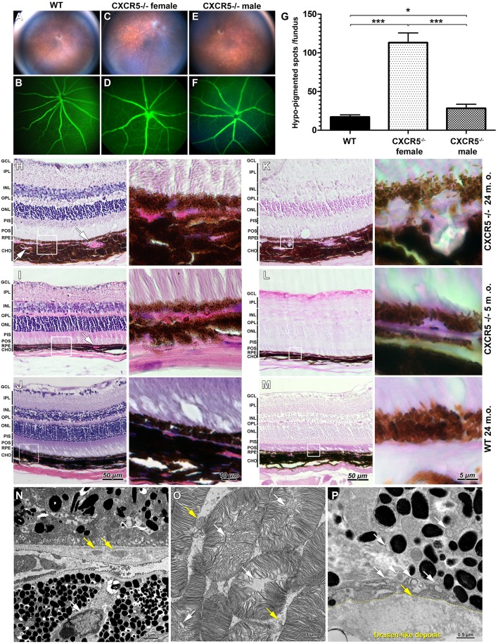
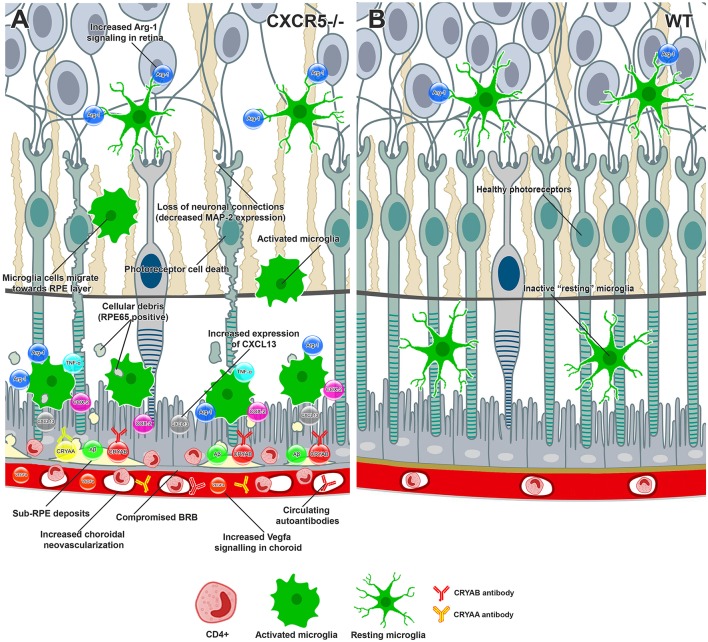
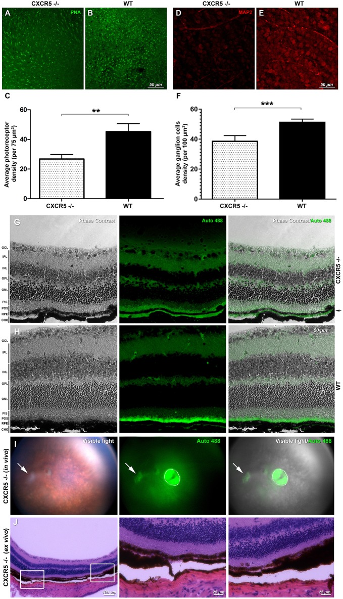
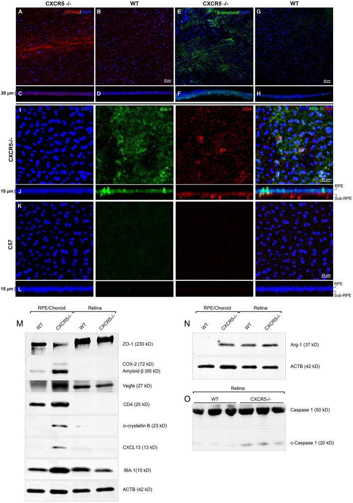
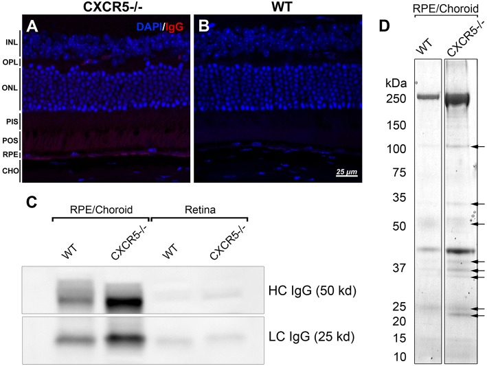
Previous research has shown that CXCR5 mice develop retinal degeneration (RD) with age, a characteristic related to age macular degeneration (AMD). RD in these mice is not well-understood, and in this study, we sought to characterize further the RD phenotype and to gain mechanistic insights into the function of CXCR5 in the retina. CXCR5 and WT control mice were used. Fundus images demonstrated a significant ( < 0.001) increase of hypo-pigmented spots in the retina of aged CXCR5 mice compared with WT control mice. PAS staining indicated localization of deposits in the sub-retinal pigment epithelia (RPE) layer. AMD-associated proteins Cryab, amyloid beta, and C3d were detected within the RPE/sub-RPE tissues by immunofluorescence (IF). In addition, western blot analysis of COX-2, Arg1, and VEGF-a revealed an increase in the signaling of these molecules within the RPE/choroid complex. Transmission electron microscopy (TEM) indicated a drusen-like structure of sub-RPE deposits with an accumulation of vacuolated cellular debris. Loss of photoreceptors was detected by peanut lectin staining and was corroborated by a reduction in MAP2 signaling. Loss of blood-retinal barrier integrity was demonstrated by a reduction of ZO-1 expression. Inflammatory cells were detected in the sub-RPE space, with an increase in IBA-1 positive microglia cells on the surface of the RPE. Mass spectrometry analysis of CXCR5 mouse RPE/choroid proteins extracts, separated by SDS-page and incubated with autologous serum, identified autoantibodies against AMD-associated proteins: Cryaa, Cryab, and Anxa2. evaluations in BV-2 cell culture indicated a significant increase in production of Arg-1 ( < 0.001) and COX-2 ( < 0.01) in the presence of anti-CXCR5 antibody when compared with Igg-treated control BV-2 cells stimulated with IL-4 and TNFα/IFNγ, respectively. Anti-CXCR5 antibody treatment without stimulating agents did not affect Arg-1 and COX-2 expression; this suggests that CXCR5 may have a regulatory role in microglia cells activation. These results indicate that with age, CXCR5 mice develop RD characterized by microglia dysfunction, increased production of CXCL13 in the RPE progressive photoreceptor, neuronal loss, and sub-RPE deposition of cellular debris, resulting in the production of immunogenic proteins and autoimmune-mediated RD.
先前的研究表明,CXCR5 小鼠随着年龄的增长会出现视网膜变性 (RD),这一特征与年龄相关性黄斑变性 (AMD) 有关。这些小鼠的 RD 机制尚不清楚,在这项研究中,我们试图进一步描述 RD 表型,并深入了解 CXCR5 在视网膜中的功能机制。我们使用了 CXCR5 和 WT 对照小鼠。眼底图像显示,与 WT 对照组相比,老龄 CXCR5 小鼠的视网膜中出现了明显(<0.001)更多的色素减退斑点。PAS 染色显示,在视网膜色素上皮 (RPE) 层下存在沉积物的定位。免疫荧光 (IF) 检测到 AMD 相关蛋白 Cryab、淀粉样 β 和 C3d 在 RPE/亚 RPE 组织中被检测到。此外,COX-2、Arg1 和 VEGF-a 的 Western blot 分析显示,这些分子在 RPE/脉络膜复合物中的信号转导增加。透射电子显微镜 (TEM) 显示,亚 RPE 沉积物呈类似 drusen 的结构,有空泡状细胞碎片堆积。花生凝集素染色检测到光感受器丧失,并通过 MAP2 信号的减少得到证实。ZO-1 表达减少表明血视网膜屏障完整性丧失。在 RPE 表面检测到亚 RPE 空间中的炎症细胞,IBA-1 阳性小胶质细胞增加。通过 SDS-page 分离并与自体血清孵育的 CXCR5 小鼠 RPE/脉络膜蛋白提取物的质谱分析,鉴定出针对 AMD 相关蛋白的自身抗体:Cryaa、Cryab 和 Anxa2。在 BV-2 细胞培养中的评估表明,与 IgG 处理的对照 BV-2 细胞相比,当用抗 CXCR5 抗体刺激时,Arg-1(<0.001)和 COX-2(<0.01)的产生显著增加,IL-4 和 TNFα/IFNγ分别刺激 BV-2 细胞。没有刺激剂的抗 CXCR5 抗体处理不会影响 Arg-1 和 COX-2 的表达;这表明 CXCR5 可能在小胶质细胞激活中起调节作用。这些结果表明,随着年龄的增长,CXCR5 小鼠会出现 RD,其特征是小胶质细胞功能障碍、RPE 中 CXCL13 的产生增加、进行性光感受器丧失、亚 RPE 细胞碎片沉积,导致免疫原性蛋白的产生和自身免疫介导的 RD。