Microbiology and Immunology Department at Medical University of South Carolina, Charleston, USA.
BMC Bioinformatics. 2019 Sep 2;20(1):448. doi: 10.1186/s12859-019-3055-3.
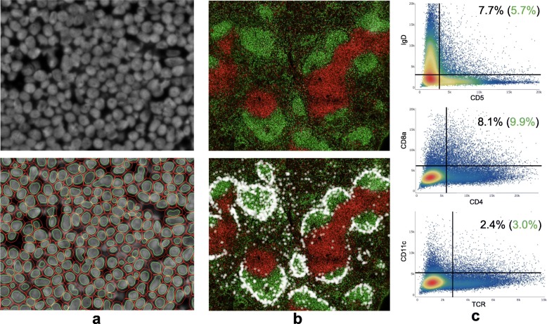
Multiplexed in-situ fluorescent imaging offers several advantages over single-cell assays that do not preserve the spatial characteristics of biological samples. This spatial information, in addition to morphological properties and extensive intracellular or surface marker profiling, comprise promising avenues for rapid advancements in the understanding of disease progression and diagnosis. As protocols for conducting such imaging experiments continue to improve, it is the intent of this study to provide and validate software for processing the large quantity of associated data in kind.
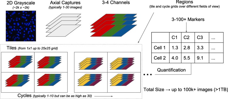
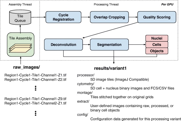
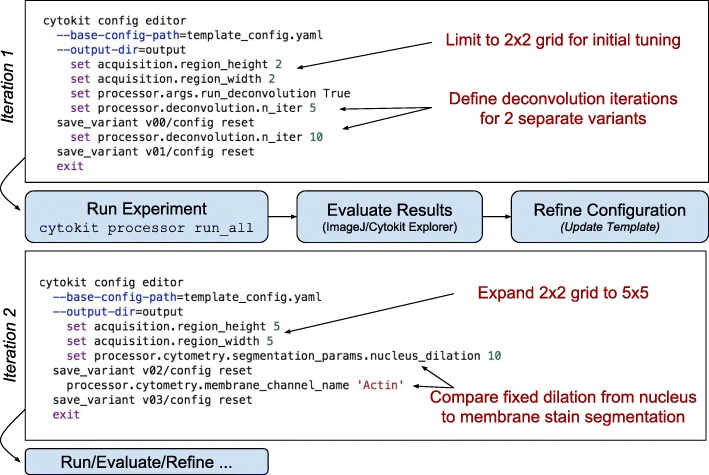
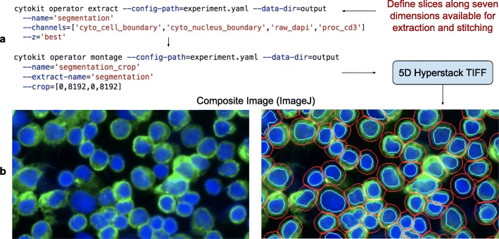
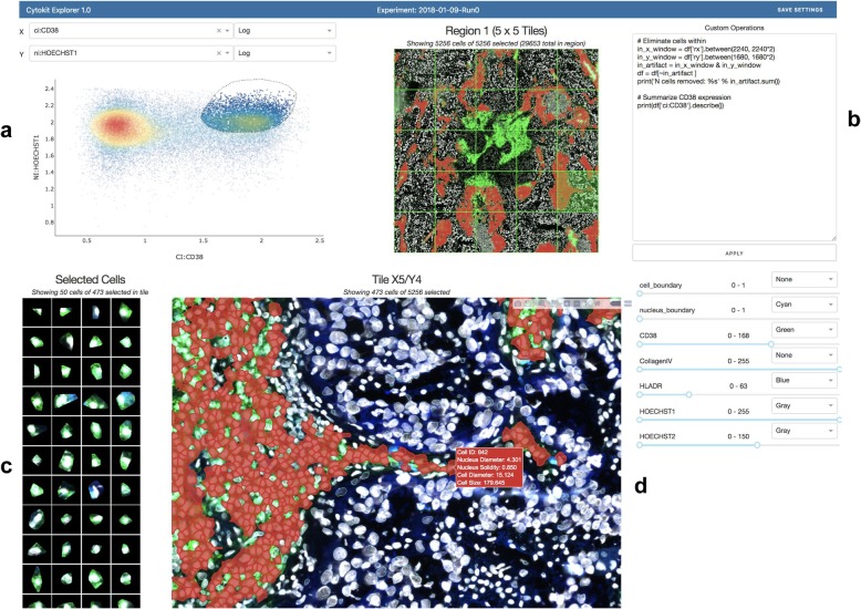
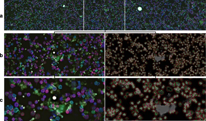
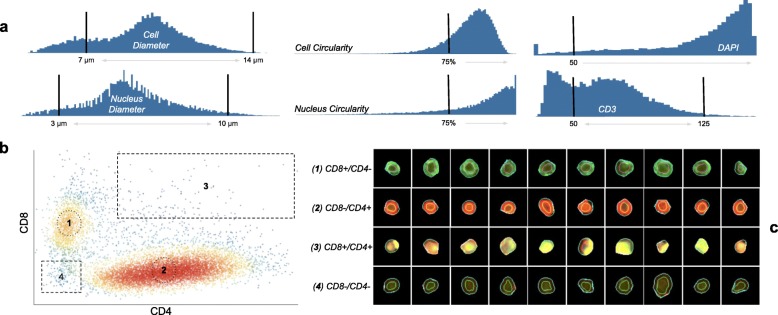
Cytokit offers (i) an end-to-end, GPU-accelerated image processing pipeline; (ii) efficient input/output (I/O) strategies for operations specific to high dimensional microscopy; and (iii) an interactive user interface for cross filtering of spatial, graphical, expression, and morphological cell properties within the 100+ GB image datasets common to multiplexed immunofluorescence. Image processing operations supported in Cytokit are generally sourced from existing deep learning models or are at least in part adapted from open source packages to run in a single or multi-GPU environment. The efficacy of these operations is demonstrated through several imaging experiments that pair Cytokit results with those from an independent but comparable assay. A further validation also demonstrates that previously published results can be reproduced from a publicly available multiplexed image dataset.
Cytokit is a collection of open source tools for quantifying and analyzing properties of individual cells in large fluorescent microscopy datasets that are often, but not necessarily, generated from multiplexed antibody labeling protocols over many fields of view or time periods. This project is best suited to bioinformaticians or other technical users that wish to analyze such data in a batch-oriented, high-throughput setting. All source code, documentation, and data generated for this article are available under the Apache License 2.0 at https://github.com/hammerlab/cytokit .
与不保留生物样本空间特征的单细胞分析相比,多重原位荧光成像具有多个优势。除了形态特征和广泛的细胞内或表面标记物分析之外,这种空间信息为快速了解疾病进展和诊断提供了有希望的途径。随着进行此类成像实验的协议不断改进,本研究旨在提供并验证用于处理相关大量数据的软件。
Cytokit 提供了 (i) 端到端、GPU 加速的图像处理管道;(ii) 针对高维显微镜特定操作的高效输入/输出 (I/O) 策略;以及 (iii) 用于在常见的多重免疫荧光 100+GB 图像数据集中交叉过滤空间、图形、表达和形态细胞特性的交互式用户界面。Cytokit 支持的图像处理操作通常源自现有深度学习模型,或者至少部分改编自开源软件包,以便在单个或多 GPU 环境中运行。通过几个成像实验证明了这些操作的有效性,这些实验将 Cytokit 的结果与来自独立但可比测定的结果进行了比较。进一步的验证还表明,可以从公开的多重图像数据集重现以前发表的结果。
Cytokit 是一组用于量化和分析大型荧光显微镜数据集中小型细胞特性的开源工具,这些数据集通常是(但不一定)由多重抗体标记方案在许多视场或时间段内生成的。该项目最适合希望在批处理、高通量环境中分析此类数据的生物信息学家或其他技术用户。本文生成的所有源代码、文档和数据均可在 Apache License 2.0 下在 https://github.com/hammerlab/cytokit 获得。