Department of Otolaryngology, Stanford University School of Medicine, Stanford, CA, United States.
Department of Microbiology and Immunology, Stanford University School of Medicine, Stanford, CA, United States.
Front Immunol. 2022 Sep 23;13:981825. doi: 10.3389/fimmu.2022.981825. eCollection 2022.
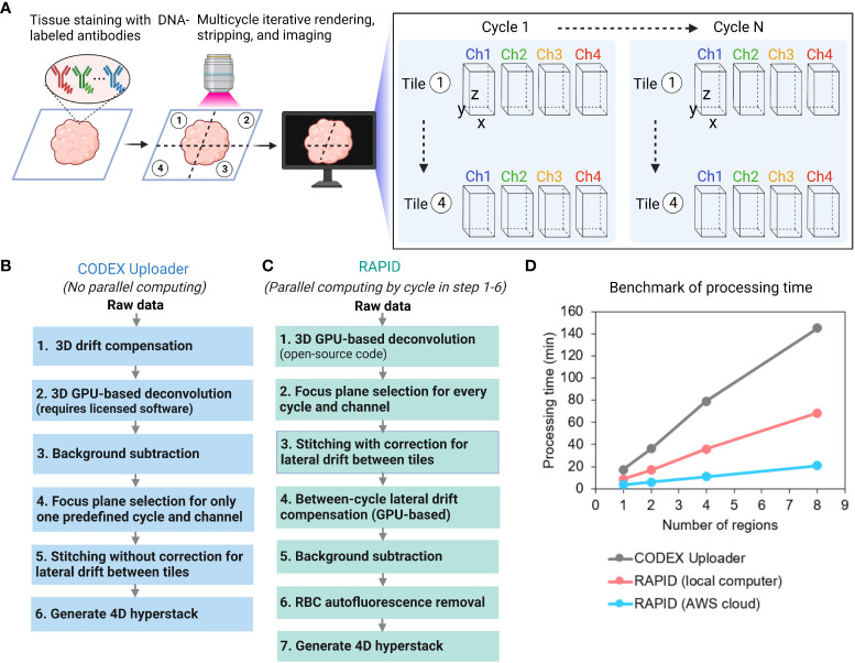
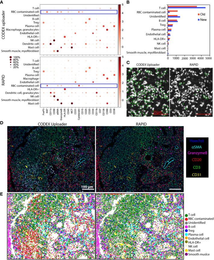
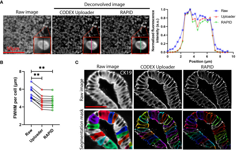
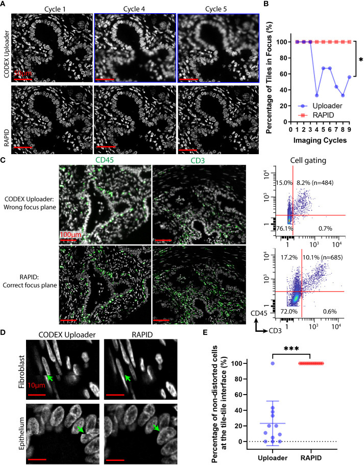
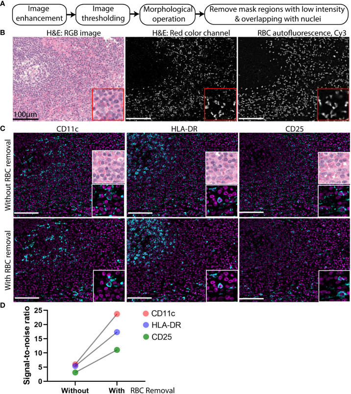
Highly multiplexed, single-cell imaging has revolutionized our understanding of spatial cellular interactions associated with health and disease. With ever-increasing numbers of antigens, region sizes, and sample sizes, multiplexed fluorescence imaging experiments routinely produce terabytes of data. Fast and accurate processing of these large-scale, high-dimensional imaging data is essential to ensure reliable segmentation and identification of cell types and for characterization of cellular neighborhoods and inference of mechanistic insights. Here, we describe RAPID, a Real-time, GPU-Accelerated Parallelized Image processing software for large-scale multiplexed fluorescence microscopy Data. RAPID deconvolves large-scale, high-dimensional fluorescence imaging data, stitches and registers images with axial and lateral drift correction, and minimizes tissue autofluorescence such as that introduced by erythrocytes. Incorporation of an open source CUDA-driven, GPU-assisted deconvolution produced results similar to fee-based commercial software. RAPID reduces data processing time and artifacts and improves image contrast and signal-to-noise compared to our previous image processing pipeline, thus providing a useful tool for accurate and robust analysis of large-scale, multiplexed, fluorescence imaging data.
高通量、单细胞成像技术极大地推动了我们对健康和疾病相关的空间细胞相互作用的理解。随着抗原数量、区域大小和样本数量的不断增加,高通量荧光成像实验通常会产生数 TB 的数据。快速准确地处理这些大规模、高维成像数据对于确保细胞类型的可靠分割和识别,以及对细胞邻域进行特征描述和推断机制见解至关重要。在这里,我们描述了 RAPID,这是一种用于大规模多色荧光显微镜数据的实时 GPU 加速并行图像处理软件。RAPID 对大规模、高维荧光成像数据进行反卷积,对带有轴向和侧向漂移校正的图像进行拼接和配准,并最小化组织自发荧光,如红细胞引起的自发荧光。结合开源的基于 CUDA 的 GPU 辅助反卷积技术,得到的结果与付费商业软件相似。与我们之前的图像处理流水线相比,RAPID 减少了数据处理时间和伪影,提高了图像对比度和信噪比,因此为大规模、多色荧光成像数据的准确和稳健分析提供了一个有用的工具。