• 文献检索
  • 文档翻译
  • 深度研究
  • 学术资讯
  • Suppr Zotero 插件Zotero 插件
  • 邀请有礼
  • 套餐&价格
  • 历史记录
应用&插件
Suppr Zotero 插件Zotero 插件浏览器插件Mac 客户端Windows 客户端微信小程序
定价
高级版会员购买积分包购买API积分包
服务
文献检索文档翻译深度研究API 文档MCP 服务
关于我们
关于 Suppr公司介绍联系我们用户协议隐私条款
关注我们

Suppr 超能文献

核心技术专利:CN118964589B侵权必究
粤ICP备2023148730 号-1Suppr @ 2026

文献检索

告别复杂PubMed语法,用中文像聊天一样搜索,搜遍4000万医学文献。AI智能推荐,让科研检索更轻松。

立即免费搜索

文件翻译

保留排版,准确专业,支持PDF/Word/PPT等文件格式,支持 12+语言互译。

免费翻译文档

深度研究

AI帮你快速写综述,25分钟生成高质量综述,智能提取关键信息,辅助科研写作。

立即免费体验

CEUS 和 MRI 联合用于诊断壶腹周围占位性病变:回顾性分析。

Combination of CEUS and MRI for the diagnosis of periampullary space-occupying lesions: a retrospective analysis.

机构信息

Department of Hepatobiliary Surgery, The Affiliated Hospital of Southwest Medical University, No. 25 Taiping Street, Jiangyang District, Luzhou, 646000, Sichuan Province, China.

Department of Nephrology, The Affiliated Hospital of Southwest Medical University, Luzhou, 646000, Sichuan Province, China.

出版信息

BMC Med Imaging. 2019 Sep 2;19(1):77. doi: 10.1186/s12880-019-0376-7.

DOI:10.1186/s12880-019-0376-7
PMID:31477041
原文链接:https://pmc.ncbi.nlm.nih.gov/articles/PMC6719361/
Abstract

BACKGROUND

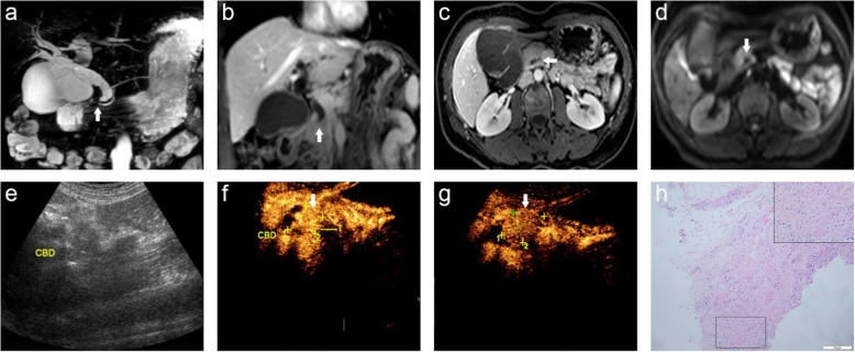
The value of magnetic resonance imaging (MRI), contrast-enhanced ultrasound (CEUS), and the combination of CEUS and MRI (CCWM) for the diagnosis of periampullary space-occupying lesions (PSOL) was investigated.

METHODS

A total of 102 patients diagnosed with PSOLs by surgery or biopsy were recruited retrospectively. The sensitivity, specificity, positive predictive value (PPV), negative predictive value (NPV), and accuracy of MRI, CEUS, and CCWM were analyzed.

RESULTS

MRI, CEUS, and CCWM allowed for the accurate detection of 91.17, 92.15, and 99.01% of PSOLs, respectively. The specificity, PPV, and accuracy of CCWM were significantly different from MRI and CEUS (p < 0.05). However, there the sensitivity and NPV were not significantly different among the three diagnostic technologies. In addition, the specificity, PPV, and accuracy were not significantly different between MRI and CEUS (all p > 0.05).

CONCLUSIONS

CCWM is valuable for differentiating benign and malignant PSOL, which provides important guiding significances for the clinic.

摘要

背景

本研究旨在探讨磁共振成像(MRI)、对比增强超声(CEUS)及 CEUS 与 MRI 联合(CCWM)对壶腹周围占位性病变(PSOL)的诊断价值。

方法

回顾性分析了 102 例经手术或活检诊断为 PSOL 的患者。分析 MRI、CEUS 和 CCWM 的灵敏度、特异度、阳性预测值(PPV)、阴性预测值(NPV)和准确率。

结果

MRI、CEUS 和 CCWM 对 PSOL 的检出率分别为 91.17%、92.15%和 99.01%。CCWM 的特异度、PPV 和准确率与 MRI 和 CEUS 相比差异有统计学意义(p<0.05)。然而,三种诊断技术之间的敏感度和 NPV 差异无统计学意义。此外,MRI 和 CEUS 的特异度、PPV 和准确率差异无统计学意义(均 p>0.05)。

结论

CCWM 对鉴别壶腹周围 PSOL 的良恶性有价值,可为临床提供重要指导意义。

https://cdn.ncbi.nlm.nih.gov/pmc/blobs/7867/6719361/c2387dbc09ab/12880_2019_376_Fig2_HTML.jpg
https://cdn.ncbi.nlm.nih.gov/pmc/blobs/7867/6719361/b67c44114cf5/12880_2019_376_Fig1_HTML.jpg
https://cdn.ncbi.nlm.nih.gov/pmc/blobs/7867/6719361/c2387dbc09ab/12880_2019_376_Fig2_HTML.jpg
https://cdn.ncbi.nlm.nih.gov/pmc/blobs/7867/6719361/b67c44114cf5/12880_2019_376_Fig1_HTML.jpg
https://cdn.ncbi.nlm.nih.gov/pmc/blobs/7867/6719361/c2387dbc09ab/12880_2019_376_Fig2_HTML.jpg

相似文献

1
Combination of CEUS and MRI for the diagnosis of periampullary space-occupying lesions: a retrospective analysis.CEUS 和 MRI 联合用于诊断壶腹周围占位性病变:回顾性分析。
BMC Med Imaging. 2019 Sep 2;19(1):77. doi: 10.1186/s12880-019-0376-7.
2
Double contrast-enhanced ultrasonography in the detection of periampullary cancer: Comparison with B-mode ultrasonography and MR imaging.双对比增强超声在壶腹周围癌检测中的应用:与B型超声和磁共振成像的比较
Eur J Radiol. 2016 Nov;85(11):1993-2000. doi: 10.1016/j.ejrad.2016.08.021. Epub 2016 Aug 28.
3
Liver Contrast-Enhanced Ultrasound Improves Detection of Liver Metastases in Patients with Pancreatic or Periampullary Cancer.肝脏对比增强超声可提高胰腺癌或壶腹周围癌患者肝转移灶的检出率。
Ultrasound Med Biol. 2015 Dec;41(12):3063-9. doi: 10.1016/j.ultrasmedbio.2015.06.019. Epub 2015 Oct 1.
4
Comparison of magnetic resonance imaging (MRI) and contrast-enhanced ultrasound (CEUS) in the evaluation of unclear solid renal lesions.磁共振成像(MRI)与对比增强超声(CEUS)在评估不明实性肾病变中的比较。
Clin Hemorheol Microcirc. 2016;64(4):757-763. doi: 10.3233/CH-168034.
5
Are a Double Duct Sign or Endoscopic Biopsies Reliable Predictors of Malignancy in Periampullary Lesions.双管征或内镜活检是否为壶腹周围病变恶性肿瘤的可靠预测指标?
Dig Surg. 2015;32(4):306-11. doi: 10.1159/000380959. Epub 2015 Jul 2.
6
Comparison of computed tomography (CT), magnetic resonance imaging (MRI) and contrast-enhanced ultrasound (CEUS) in the evaluation of unclear renal lesions.计算机断层扫描(CT)、磁共振成像(MRI)和对比增强超声(CEUS)在评估不明原因肾脏病变中的比较。
Rofo. 2020 Nov;192(11):1053-1059. doi: 10.1055/a-1127-3371. Epub 2020 Apr 15.
7
LI-RADS-CEUS - Proposal for a Contrast-Enhanced Ultrasound Algorithm for the Diagnosis of Hepatocellular Carcinoma in High-Risk Populations.肝脏影像报告和数据系统-超声造影 - 关于高危人群肝细胞癌诊断的超声造影算法提案
Ultraschall Med. 2016 Dec;37(6):627-634. doi: 10.1055/s-0042-112221. Epub 2016 Aug 3.
8
MRI combined with MR cholangiopancreatography versus helical CT in the evaluation of patients with suspected periampullary tumors: a prospective comparative study.磁共振成像联合磁共振胰胆管造影与螺旋CT在疑似壶腹周围肿瘤患者评估中的比较:一项前瞻性对照研究
Acta Radiol. 2005 Feb;46(1):16-27. doi: 10.1080/02841850510016018.
9
Differential diagnosis of periampullary carcinomas: comparison of CT with negative-contrast CT cholangiopancreatography versus MRI with MR cholangiopancreatography.壶腹周围癌的鉴别诊断:CT与阴性对比剂CT胰胆管造影术对比MRI与磁共振胰胆管造影术的比较
Abdom Imaging. 2014 Jun;39(3):506-17. doi: 10.1007/s00261-014-0085-1.
10
Diagnostic efficacy of contrast-enhanced ultrasound for breast lesions of different sizes: a comparative study with magnetic resonance imaging.不同大小乳腺病变超声造影诊断效能的对比研究:与磁共振成像的对照研究。
Br J Radiol. 2020 Jun;93(1110):20190932. doi: 10.1259/bjr.20190932. Epub 2020 Apr 7.

引用本文的文献

1
Application of imaging technology for the diagnosis of malignancy in the pancreaticobiliary duodenal junction (Review).成像技术在胰胆管十二指肠交界处恶性肿瘤诊断中的应用(综述)
Oncol Lett. 2024 Oct 8;28(6):596. doi: 10.3892/ol.2024.14729. eCollection 2024 Dec.
2
Identifying Periampullary Regions in MRI Images Using Deep Learning.使用深度学习在MRI图像中识别壶腹周围区域。
Front Oncol. 2021 May 28;11:674579. doi: 10.3389/fonc.2021.674579. eCollection 2021.

本文引用的文献

1
Negative Predictive Value of Contrast-Enhanced Ultrasound in Differentiating Avascular Solid-Appearing From Vascularized Masses: A Retrospective Consecutive Study.超声造影在鉴别无血管实性表现与血管化肿块中的阴性预测价值:一项回顾性连续研究。
J Ultrasound Med. 2018 Dec;37(12):2935-2942. doi: 10.1002/jum.14659. Epub 2018 May 15.
2
Diagnostic Accuracy of a 64-Slice Multi-Detector CT Scan in the Preoperative Evaluation of Periampullary Neoplasms.64层多排螺旋CT扫描在壶腹周围肿瘤术前评估中的诊断准确性
J Clin Med. 2018 Apr 27;7(5):91. doi: 10.3390/jcm7050091.
3
Prognostic Factors and the Role of Adjuvant Treatment in Periampullary Carcinoma: a Single-Centre Experience of 95 Patients.
壶腹周围癌的预后因素及辅助治疗的作用:95例患者的单中心经验
J Gastrointest Cancer. 2019 Sep;50(3):361-369. doi: 10.1007/s12029-018-0058-7.
4
Double contrast-enhanced ultrasonography in the detection of periampullary cancer: Comparison with B-mode ultrasonography and MR imaging.双对比增强超声在壶腹周围癌检测中的应用:与B型超声和磁共振成像的比较
Eur J Radiol. 2016 Nov;85(11):1993-2000. doi: 10.1016/j.ejrad.2016.08.021. Epub 2016 Aug 28.
5
Differentiation grade for extrahepatic bile duct adenocarcinoma: Assessed by diffusion-weighted imaging at 3.0-T MR.肝外胆管腺癌的分化程度:通过3.0-T磁共振成像的扩散加权成像评估。
Eur J Radiol. 2016 Nov;85(11):1980-1986. doi: 10.1016/j.ejrad.2016.09.004. Epub 2016 Sep 9.
6
Differentiation of pancreatic carcinoma and mass-forming focal pancreatitis: qualitative and quantitative assessment by dynamic contrast-enhanced MRI combined with diffusion-weighted imaging.胰腺癌与肿块型局灶性胰腺炎的鉴别:动态对比增强磁共振成像联合扩散加权成像的定性与定量评估
Oncotarget. 2017 Jan 3;8(1):1744-1759. doi: 10.18632/oncotarget.12120.
7
Liver Contrast-Enhanced Ultrasound Improves Detection of Liver Metastases in Patients with Pancreatic or Periampullary Cancer.肝脏对比增强超声可提高胰腺癌或壶腹周围癌患者肝转移灶的检出率。
Ultrasound Med Biol. 2015 Dec;41(12):3063-9. doi: 10.1016/j.ultrasmedbio.2015.06.019. Epub 2015 Oct 1.
8
Accuracy of combined dynamic contrast-enhanced magnetic resonance imaging and diffusion-weighted imaging for breast cancer detection: a meta-analysis.联合动态对比增强磁共振成像和扩散加权成像检测乳腺癌的准确性:一项荟萃分析。
Acta Radiol. 2016 Jun;57(6):651-60. doi: 10.1177/0284185115597265. Epub 2015 Aug 13.
9
Prognostic factors affecting survival after pancreaticoduodenectomy for pancreatic adenocarcinoma (single center experience).影响胰腺腺癌胰十二指肠切除术后生存的预后因素(单中心经验)
Hepatogastroenterology. 2014 Jul-Aug;61(133):1426-38.
10
Differentiation of noncalculous periampullary obstruction: comparison of CT with negative-contrast CT cholangiopancreatography versus MRI with MR cholangiopancreatography.非结石性胰胆管壶腹周围梗阻的鉴别诊断:CT 与阴性对比 CT 胆胰管成像和 MRI 与 MR 胆胰管成像的比较。
Eur Radiol. 2015 Feb;25(2):391-401. doi: 10.1007/s00330-014-3430-4. Epub 2014 Sep 24.