• 文献检索
  • 文档翻译
  • 深度研究
  • 学术资讯
  • Suppr Zotero 插件Zotero 插件
  • 邀请有礼
  • 套餐&价格
  • 历史记录
应用&插件
Suppr Zotero 插件Zotero 插件浏览器插件Mac 客户端Windows 客户端微信小程序
定价
高级版会员购买积分包购买API积分包
服务
文献检索文档翻译深度研究API 文档MCP 服务
关于我们
关于 Suppr公司介绍联系我们用户协议隐私条款
关注我们

Suppr 超能文献

核心技术专利:CN118964589B侵权必究
粤ICP备2023148730 号-1Suppr @ 2026

文献检索

告别复杂PubMed语法,用中文像聊天一样搜索,搜遍4000万医学文献。AI智能推荐,让科研检索更轻松。

立即免费搜索

文件翻译

保留排版,准确专业,支持PDF/Word/PPT等文件格式,支持 12+语言互译。

免费翻译文档

深度研究

AI帮你快速写综述,25分钟生成高质量综述,智能提取关键信息,辅助科研写作。

立即免费体验

用于肺部CT图像预筛查的人工智能算法测试的统计学考量

Statistical considerations for testing an AI algorithm used for prescreening lung CT images.

作者信息

Obuchowski Nancy A, Bullen Jennifer A

机构信息

Quantitative Health Sciences /JJN3, Cleveland Clinic Foundation, 9500 Euclid Ave, Cleveland, OH, 44195, USA.

出版信息

Contemp Clin Trials Commun. 2019 Aug 22;16:100434. doi: 10.1016/j.conctc.2019.100434. eCollection 2019 Dec.

DOI:10.1016/j.conctc.2019.100434
PMID:31485545
原文链接:https://pmc.ncbi.nlm.nih.gov/articles/PMC6717063/
Abstract

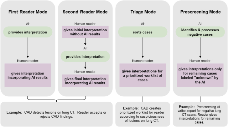
Artificial intelligence, as applied to medical images to detect, rule out, diagnose, and stage disease, has seen enormous growth over the last few years. There are multiple use cases of AI algorithms in medical imaging: first-reader (or concurrent) mode, second-reader mode, triage mode, and more recently prescreening mode as when an AI algorithm is applied to the worklist of images to identify obvious negative cases so that human readers do not need to review them and can focus on interpreting the remaining cases. In this paper we describe the statistical considerations for designing a study to test a new AI prescreening algorithm for identifying normal lung cancer screening CTs. We contrast agreement vs. accuracy studies, and retrospective vs. prospective designs. We evaluate various test performance metrics with respect to their sensitivity to changes in the AI algorithm's performance, as well as to shifts in reader behavior to a revised worklist. We consider sample size requirements for testing the AI prescreening algorithm.

摘要

在过去几年中,应用于医学图像以检测、排除、诊断疾病及确定疾病分期的人工智能技术取得了巨大发展。人工智能算法在医学成像中有多种应用场景:初读(或同步)模式、复阅模式、分诊模式,以及最近出现的预筛查模式,即当将人工智能算法应用于图像工作列表以识别明显的阴性病例时,人类阅片者无需查看这些病例,而是可以专注于解读其余病例。在本文中,我们描述了设计一项研究的统计学考量,该研究旨在测试一种用于识别正常肺癌筛查CT的新型人工智能预筛查算法。我们对比了一致性研究与准确性研究,以及回顾性设计与前瞻性设计。我们评估了各种测试性能指标,考量它们对人工智能算法性能变化的敏感性,以及对阅片者行为向修订后的工作列表转变的敏感性。我们还考虑了测试人工智能预筛查算法所需的样本量。

https://cdn.ncbi.nlm.nih.gov/pmc/blobs/92b7/6717063/25cf1b1a4784/gr5.jpg
https://cdn.ncbi.nlm.nih.gov/pmc/blobs/92b7/6717063/450421c80365/gr1.jpg
https://cdn.ncbi.nlm.nih.gov/pmc/blobs/92b7/6717063/825075b7f912/gr2.jpg
https://cdn.ncbi.nlm.nih.gov/pmc/blobs/92b7/6717063/72fe0f42707b/gr3.jpg
https://cdn.ncbi.nlm.nih.gov/pmc/blobs/92b7/6717063/b2776606cfd0/gr4.jpg
https://cdn.ncbi.nlm.nih.gov/pmc/blobs/92b7/6717063/25cf1b1a4784/gr5.jpg

相似文献

1
Statistical considerations for testing an AI algorithm used for prescreening lung CT images.用于肺部CT图像预筛查的人工智能算法测试的统计学考量
Contemp Clin Trials Commun. 2019 Aug 22;16:100434. doi: 10.1016/j.conctc.2019.100434. eCollection 2019 Dec.
2
Artificial Intelligence-Based Identification of Normal Chest Radiographs: A Simulation Study in a Multicenter Health Screening Cohort.基于人工智能的正常胸部 X 光片识别:多中心健康筛查队列中的模拟研究。
Korean J Radiol. 2022 Oct;23(10):1009-1018. doi: 10.3348/kjr.2022.0189.
3
Diagnostic study on clinical feasibility of an AI-based diagnostic system as a second reader on mobile CT images: a preliminary result.基于人工智能的诊断系统作为移动CT图像第二阅片者的临床可行性诊断研究:初步结果
Ann Transl Med. 2022 Jun;10(12):668. doi: 10.21037/atm-22-2157.
4
Artificial Intelligence Tool for Detection and Worklist Prioritization Reduces Time to Diagnosis of Incidental Pulmonary Embolism at CT.用于检测和工作列表优先级排序的人工智能工具可减少CT诊断偶然发现的肺栓塞的时间。
Radiol Cardiothorac Imaging. 2023 Apr 20;5(2):e220163. doi: 10.1148/ryct.220163. eCollection 2023 Apr.
5
Real-world testing of an artificial intelligence algorithm for the analysis of chest X-rays in primary care settings.用于基层医疗环境中胸部X光分析的人工智能算法的真实世界测试。
Sci Rep. 2024 Mar 3;14(1):5199. doi: 10.1038/s41598-024-55792-1.
6
Using Artificial Intelligence to Revise ACR TI-RADS Risk Stratification of Thyroid Nodules: Diagnostic Accuracy and Utility.使用人工智能修订甲状腺结节 ACR TI-RADS 风险分层:诊断准确性和实用性。
Radiology. 2019 Jul;292(1):112-119. doi: 10.1148/radiol.2019182128. Epub 2019 May 21.
7
Improving Clinical Trial Participant Prescreening With Artificial Intelligence (AI): A Comparison of the Results of AI-Assisted vs Standard Methods in 3 Oncology Trials.利用人工智能(AI)改进临床试验参与者的预筛选:3 项肿瘤学试验中 AI 辅助与标准方法结果的比较。
Ther Innov Regul Sci. 2020 Jan;54(1):69-74. doi: 10.1007/s43441-019-00030-4. Epub 2020 Jan 6.
8
Detection and Diagnosis of Breast Cancer Using Artificial Intelligence Based assessment of Maximum Intensity Projection Dynamic Contrast-Enhanced Magnetic Resonance Images.基于人工智能评估最大强度投影动态对比增强磁共振图像的乳腺癌检测与诊断
Diagnostics (Basel). 2020 May 20;10(5):330. doi: 10.3390/diagnostics10050330.
9
The development an artificial intelligence algorithm for early sepsis diagnosis in the intensive care unit.开发一种用于重症监护病房早期脓毒症诊断的人工智能算法。
Int J Med Inform. 2020 Sep;141:104176. doi: 10.1016/j.ijmedinf.2020.104176. Epub 2020 May 21.
10
Improved Cancer Detection Using Artificial Intelligence: a Retrospective Evaluation of Missed Cancers on Mammography.利用人工智能提高癌症检测率:对乳腺 X 光摄影术漏诊癌症的回顾性评估。
J Digit Imaging. 2019 Aug;32(4):625-637. doi: 10.1007/s10278-019-00192-5.

引用本文的文献

1
A Thorough Review of the Clinical Applications of Artificial Intelligence in Lung Cancer.人工智能在肺癌临床应用的全面综述
Cancers (Basel). 2025 Mar 4;17(5):882. doi: 10.3390/cancers17050882.
2
An Assessment of Deep Learning's Impact on General Dentists' Ability to Detect Alveolar Bone Loss in 2D Intraoral Radiographs.深度学习对普通牙医在二维口腔内X光片中检测牙槽骨丧失能力的影响评估
Diagnostics (Basel). 2025 Feb 14;15(4):467. doi: 10.3390/diagnostics15040467.
3
Improving mammography interpretation for both novice and experienced readers: a comparative study of two commercial artificial intelligence software.

本文引用的文献

1
Design Characteristics of Studies Reporting the Performance of Artificial Intelligence Algorithms for Diagnostic Analysis of Medical Images: Results from Recently Published Papers.研究报告用于医学图像诊断分析的人工智能算法性能的设计特点:近期发表论文的结果。
Korean J Radiol. 2019 Mar;20(3):405-410. doi: 10.3348/kjr.2019.0025.
2
Deep-learning-assisted diagnosis for knee magnetic resonance imaging: Development and retrospective validation of MRNet.深度学习辅助膝关节磁共振成像诊断:MRNet 的开发和回顾性验证。
PLoS Med. 2018 Nov 27;15(11):e1002699. doi: 10.1371/journal.pmed.1002699. eCollection 2018 Nov.
3
Detection of Breast Cancer with Mammography: Effect of an Artificial Intelligence Support System.
提高新手和有经验读者的乳腺 X 线摄影判读能力:两种商业人工智能软件的对比研究。
Eur Radiol. 2024 Jun;34(6):3924-3934. doi: 10.1007/s00330-023-10422-8. Epub 2023 Nov 8.
4
Effects of a comprehensive brain computed tomography deep learning model on radiologist detection accuracy.基于深度学习的脑 CT 综合模型对放射科医生检测准确率的影响。
Eur Radiol. 2024 Feb;34(2):810-822. doi: 10.1007/s00330-023-10074-8. Epub 2023 Aug 22.
5
"Shortcuts" Causing Bias in Radiology Artificial Intelligence: Causes, Evaluation, and Mitigation.“捷径”导致放射科人工智能产生偏见:原因、评估和缓解。
J Am Coll Radiol. 2023 Sep;20(9):842-851. doi: 10.1016/j.jacr.2023.06.025. Epub 2023 Jul 27.
6
Improving the diagnosis of acute ischemic stroke on non-contrast CT using deep learning: a multicenter study.利用深度学习提高非增强CT对急性缺血性卒中的诊断:一项多中心研究。
Insights Imaging. 2022 Dec 6;13(1):184. doi: 10.1186/s13244-022-01331-3.
7
Possible Bias in Supervised Deep Learning Algorithms for CT Lung Nodule Detection and Classification.用于CT肺结节检测与分类的监督深度学习算法中的潜在偏差
Cancers (Basel). 2022 Aug 10;14(16):3867. doi: 10.3390/cancers14163867.
8
Using Occlusion-Based Saliency Maps to Explain an Artificial Intelligence Tool in Lung Cancer Screening: Agreement Between Radiologists, Labels, and Visual Prompts.基于掩蔽的显著图在肺癌筛查人工智能工具解释中的应用:放射科医生、标注者和视觉提示之间的一致性。
J Digit Imaging. 2022 Oct;35(5):1164-1175. doi: 10.1007/s10278-022-00631-w. Epub 2022 Apr 28.
9
Do comprehensive deep learning algorithms suffer from hidden stratification? A retrospective study on pneumothorax detection in chest radiography.综合深度学习算法是否存在隐藏分层问题?一项关于胸部 X 光片中气胸检测的回顾性研究。
BMJ Open. 2021 Dec 7;11(12):e053024. doi: 10.1136/bmjopen-2021-053024.
10
Artificial intelligence for detection and characterization of pulmonary nodules in lung cancer CT screening: ready for practice?人工智能在肺癌CT筛查中检测和表征肺结节:准备好应用于临床了吗?
Transl Lung Cancer Res. 2021 May;10(5):2378-2388. doi: 10.21037/tlcr-2020-lcs-06.
乳腺 X 线摄影术检测乳腺癌:人工智能支持系统的效果。
Radiology. 2019 Feb;290(2):305-314. doi: 10.1148/radiol.2018181371. Epub 2018 Nov 20.
4
Machine learning analyses can differentiate meningioma grade by features on magnetic resonance imaging.机器学习分析可以通过磁共振成像上的特征来区分脑膜瘤的级别。
Neurosurg Focus. 2018 Nov 1;45(5):E4. doi: 10.3171/2018.8.FOCUS18191.
5
Demystification of AI-driven medical image interpretation: past, present and future.人工智能驱动的医学图像解读的解密:过去、现在和未来。
Eur Radiol. 2019 Mar;29(3):1616-1624. doi: 10.1007/s00330-018-5674-x. Epub 2018 Aug 13.
6
Data Analysis Strategies in Medical Imaging.医学影像学中的数据分析策略。
Clin Cancer Res. 2018 Aug 1;24(15):3492-3499. doi: 10.1158/1078-0432.CCR-18-0385. Epub 2018 Mar 26.
7
Artificial intelligence in healthcare: past, present and future.人工智能在医疗保健中的应用:过去、现在和未来。
Stroke Vasc Neurol. 2017 Jun 21;2(4):230-243. doi: 10.1136/svn-2017-000101. eCollection 2017 Dec.
8
Methodologic Guide for Evaluating Clinical Performance and Effect of Artificial Intelligence Technology for Medical Diagnosis and Prediction.医学诊断和预测人工智能技术临床效能评估的方法学指南
Radiology. 2018 Mar;286(3):800-809. doi: 10.1148/radiol.2017171920. Epub 2018 Jan 8.
9
Computer-aided detection improves detection of pulmonary nodules in chest radiographs beyond the support by bone-suppressed images.计算机辅助检测可提高胸部 X 线摄影中肺结节的检出率,优于骨抑制图像的支持。
Radiology. 2014 Jul;272(1):252-61. doi: 10.1148/radiol.14131315. Epub 2014 Mar 12.
10
Results of initial low-dose computed tomographic screening for lung cancer.肺癌初始低剂量计算机断层扫描筛查结果。
N Engl J Med. 2013 May 23;368(21):1980-91. doi: 10.1056/NEJMoa1209120.