• 文献检索
  • 文档翻译
  • 深度研究
  • 学术资讯
  • Suppr Zotero 插件Zotero 插件
  • 邀请有礼
  • 套餐&价格
  • 历史记录
应用&插件
Suppr Zotero 插件Zotero 插件浏览器插件Mac 客户端Windows 客户端微信小程序
定价
高级版会员购买积分包购买API积分包
服务
文献检索文档翻译深度研究API 文档MCP 服务
关于我们
关于 Suppr公司介绍联系我们用户协议隐私条款
关注我们

Suppr 超能文献

核心技术专利:CN118964589B侵权必究
粤ICP备2023148730 号-1Suppr @ 2026

文献检索

告别复杂PubMed语法,用中文像聊天一样搜索,搜遍4000万医学文献。AI智能推荐,让科研检索更轻松。

立即免费搜索

文件翻译

保留排版,准确专业,支持PDF/Word/PPT等文件格式,支持 12+语言互译。

免费翻译文档

深度研究

AI帮你快速写综述,25分钟生成高质量综述,智能提取关键信息,辅助科研写作。

立即免费体验

基于人工智能评估最大强度投影动态对比增强磁共振图像的乳腺癌检测与诊断

Detection and Diagnosis of Breast Cancer Using Artificial Intelligence Based assessment of Maximum Intensity Projection Dynamic Contrast-Enhanced Magnetic Resonance Images.

作者信息

Adachi Mio, Fujioka Tomoyuki, Mori Mio, Kubota Kazunori, Kikuchi Yuka, Xiaotong Wu, Oyama Jun, Kimura Koichiro, Oda Goshi, Nakagawa Tsuyoshi, Uetake Hiroyuki, Tateishi Ukihide

机构信息

Department of Surgery, Breast Surgery, Tokyo Medical and Dental University, Tokyo 113-8510, Japan.

Department of Diagnostic Radiology, Tokyo Medical and Dental University, Tokyo 113-8510, Japan.

出版信息

Diagnostics (Basel). 2020 May 20;10(5):330. doi: 10.3390/diagnostics10050330.

DOI:10.3390/diagnostics10050330
PMID:32443922
原文链接:https://pmc.ncbi.nlm.nih.gov/articles/PMC7277981/
Abstract

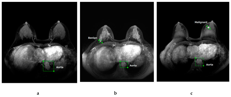
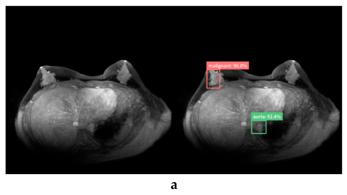
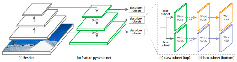
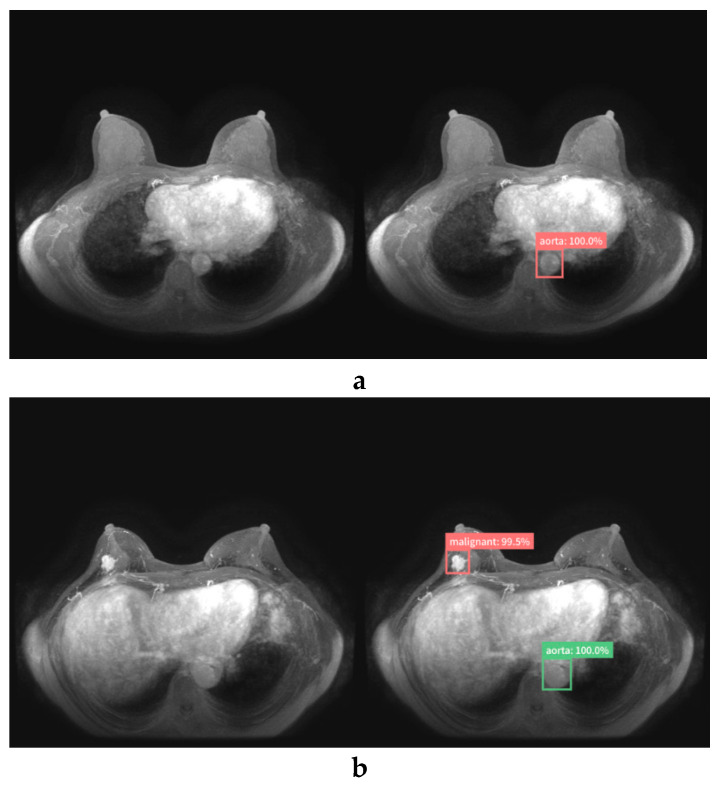
We aimed to evaluate an artificial intelligence (AI) system that can detect and diagnose lesions of maximum intensity projection (MIP) in dynamic contrast-enhanced (DCE) breast magnetic resonance imaging (MRI). We retrospectively gathered MIPs of DCE breast MRI for training and validation data from 30 and 7 normal individuals, 49 and 20 benign cases, and 135 and 45 malignant cases, respectively. Breast lesions were indicated with a bounding box and labeled as benign or malignant by a radiologist, while the AI system was trained to detect and calculate possibilities of malignancy using RetinaNet. The AI system was analyzed using test sets of 13 normal, 20 benign, and 52 malignant cases. Four human readers also scored these test data with and without the assistance of the AI system for the possibility of a malignancy in each breast. Sensitivity, specificity, and area under the receiver operating characteristic curve (AUC) were 0.926, 0.828, and 0.925 for the AI system; 0.847, 0.841, and 0.884 for human readers without AI; and 0.889, 0.823, and 0.899 for human readers with AI using a cutoff value of 2%, respectively. The AI system showed better diagnostic performance compared to the human readers ( = 0.002), and because of the increased performance of human readers with the assistance of the AI system, the AUC of human readers was significantly higher with than without the AI system ( = 0.039). Our AI system showed a high performance ability in detecting and diagnosing lesions in MIPs of DCE breast MRI and increased the diagnostic performance of human readers.

摘要

我们旨在评估一种人工智能(AI)系统,该系统能够在动态对比增强(DCE)乳腺磁共振成像(MRI)中检测和诊断最大强度投影(MIP)的病变。我们回顾性收集了DCE乳腺MRI的MIP图像,分别用于30名正常个体和7名正常个体、49例良性病例和20例良性病例、135例恶性病例和45例恶性病例的训练和验证数据。乳腺病变用边界框表示,并由放射科医生标记为良性或恶性,而AI系统则使用RetinaNet进行训练,以检测和计算恶性可能性。使用13例正常、20例良性和52例恶性病例的测试集对AI系统进行分析。四位人类读者也对这些测试数据进行了评分,分别在有无AI系统辅助的情况下评估每个乳腺发生恶性病变的可能性。AI系统的灵敏度、特异度和受试者操作特征曲线下面积(AUC)分别为0.926、0.828和0.925;无AI辅助的人类读者分别为0.847、0.841和0.884;使用2%的临界值时,有AI辅助的人类读者分别为0.889、0.823和0.899。与人类读者相比,AI系统表现出更好的诊断性能(P = 0.002),并且由于在AI系统辅助下人类读者的性能有所提高,有AI系统时人类读者的AUC显著高于无AI系统时(P = 0.039)。我们的AI系统在检测和诊断DCE乳腺MRI的MIP病变方面表现出高性能,并提高了人类读者的诊断性能。

https://cdn.ncbi.nlm.nih.gov/pmc/blobs/b972/7277981/0de1eea77ff6/diagnostics-10-00330-g006a.jpg
https://cdn.ncbi.nlm.nih.gov/pmc/blobs/b972/7277981/51638f12b6b0/diagnostics-10-00330-g001.jpg
https://cdn.ncbi.nlm.nih.gov/pmc/blobs/b972/7277981/f70a745b864c/diagnostics-10-00330-g002.jpg
https://cdn.ncbi.nlm.nih.gov/pmc/blobs/b972/7277981/5e2a9f91d7a1/diagnostics-10-00330-g003.jpg
https://cdn.ncbi.nlm.nih.gov/pmc/blobs/b972/7277981/0ab48c4d825a/diagnostics-10-00330-g004.jpg
https://cdn.ncbi.nlm.nih.gov/pmc/blobs/b972/7277981/b3ea4dce2cc3/diagnostics-10-00330-g005a.jpg
https://cdn.ncbi.nlm.nih.gov/pmc/blobs/b972/7277981/0de1eea77ff6/diagnostics-10-00330-g006a.jpg

相似文献

1
Detection and Diagnosis of Breast Cancer Using Artificial Intelligence Based assessment of Maximum Intensity Projection Dynamic Contrast-Enhanced Magnetic Resonance Images.基于人工智能评估最大强度投影动态对比增强磁共振图像的乳腺癌检测与诊断
Diagnostics (Basel). 2020 May 20;10(5):330. doi: 10.3390/diagnostics10050330.
2
Deep-learning approach with convolutional neural network for classification of maximum intensity projections of dynamic contrast-enhanced breast magnetic resonance imaging.基于卷积神经网络的深度学习方法在动态对比增强乳腺磁共振成像最大强度投影中的分类。
Magn Reson Imaging. 2021 Jan;75:1-8. doi: 10.1016/j.mri.2020.10.003. Epub 2020 Oct 10.
3
An artificial intelligence system using maximum intensity projection MR images facilitates classification of non-mass enhancement breast lesions.基于最大密度投影磁共振图像的人工智能系统有助于非肿块样强化乳腺病变的分类。
Eur Radiol. 2022 Jul;32(7):4857-4867. doi: 10.1007/s00330-022-08553-5. Epub 2022 Mar 8.
4
Independent validation of machine learning in diagnosing breast Cancer on magnetic resonance imaging within a single institution.在单家医疗机构内使用机器学习对乳腺磁共振成像进行独立验证诊断乳腺癌。
Cancer Imaging. 2019 Sep 18;19(1):64. doi: 10.1186/s40644-019-0252-2.
5
Characterization of spatiotemporal changes for the classification of dynamic contrast-enhanced magnetic-resonance breast lesions.动态对比增强磁共振乳腺病变分类的时空变化特征。
Artif Intell Med. 2013 Jun;58(2):101-14. doi: 10.1016/j.artmed.2013.03.002. Epub 2013 Mar 30.
6
Artificial Intelligence-Based Classification of Breast Lesions Imaged With a Multiparametric Breast MRI Protocol With Ultrafast DCE-MRI, T2, and DWI.基于人工智能的多参数乳腺 MRI 协议(包括超快速 DCE-MRI、T2 和 DWI)成像的乳腺病变分类。
Invest Radiol. 2019 Jun;54(6):325-332. doi: 10.1097/RLI.0000000000000544.
7
Improved Classification of Benign and Malignant Breast Lesions Using Deep Feature Maximum Intensity Projection MRI in Breast Cancer Diagnosis Using Dynamic Contrast-enhanced MRI.在使用动态对比增强磁共振成像的乳腺癌诊断中,利用深度特征最大强度投影磁共振成像改进乳腺良恶性病变的分类
Radiol Artif Intell. 2021 Feb 24;3(3):e200159. doi: 10.1148/ryai.2021200159. eCollection 2021 May.
8
Improving the Performance of Radiologists Using Artificial Intelligence-Based Detection Support Software for Mammography: A Multi-Reader Study.利用基于人工智能的乳腺 X 线摄影检测支持软件提高放射科医生的工作表现:一项多读者研究。
Korean J Radiol. 2022 May;23(5):505-516. doi: 10.3348/kjr.2021.0476. Epub 2022 Apr 4.
9
Differentiation of breast lesions on dynamic contrast-enhanced magnetic resonance imaging (DCE-MRI) using deep transfer learning based on DenseNet201.基于 DenseNet201 的深度迁移学习在动态对比增强磁共振成像(DCE-MRI)上对乳腺病变的鉴别诊断。
Medicine (Baltimore). 2022 Nov 11;101(45):e31214. doi: 10.1097/MD.0000000000031214.
10
Automated artifact detection in abbreviated dynamic contrast-enhanced (DCE) MRI-derived maximum intensity projections (MIPs) of the breast.自动检测缩短动态对比增强磁共振成像(DCE-MRI)衍生乳腺最大强度投影(MIP)中的伪影。
Eur Radiol. 2022 Sep;32(9):5997-6007. doi: 10.1007/s00330-022-08626-5. Epub 2022 Apr 2.

引用本文的文献

1
Predicting Very Early-Stage Breast Cancer in BI-RADS 3 Lesions of Large Population with Deep Learning.利用深度学习预测大量人群BI-RADS 3类病变中的极早期乳腺癌。
J Imaging. 2025 Jul 15;11(7):240. doi: 10.3390/jimaging11070240.
2
Application of Machine Learning to Breast MR Imaging.机器学习在乳腺磁共振成像中的应用。
Magn Reson Med Sci. 2025 Jul 1;24(3):279-299. doi: 10.2463/mrms.rev.2025-0021. Epub 2025 Jun 14.
3
Deep learning-based breast cancer diagnosis in breast MRI: systematic review and meta-analysis.基于深度学习的乳腺MRI乳腺癌诊断:系统评价与荟萃分析

本文引用的文献

1
A machine learning approach on multiscale texture analysis for breast microcalcification diagnosis.基于多尺度纹理分析的机器学习方法在乳腺微钙化诊断中的应用。
BMC Bioinformatics. 2020 Mar 11;21(Suppl 2):91. doi: 10.1186/s12859-020-3358-4.
2
Deep Learning in Medical Imaging.医学成像中的深度学习
Neurospine. 2019 Dec;16(4):657-668. doi: 10.14245/ns.1938396.198. Epub 2019 Dec 31.
3
Breast Ultrasound Image Synthesis using Deep Convolutional Generative Adversarial Networks.使用深度卷积生成对抗网络的乳腺超声图像合成
Eur Radiol. 2025 Feb 5. doi: 10.1007/s00330-025-11406-6.
4
Deep learning-based object detection algorithms in medical imaging: Systematic review.医学成像中基于深度学习的目标检测算法:系统综述
Heliyon. 2024 Dec 11;11(1):e41137. doi: 10.1016/j.heliyon.2024.e41137. eCollection 2025 Jan 15.
5
Empowering cancer prevention with AI: unlocking new frontiers in prediction, diagnosis, and intervention.利用人工智能助力癌症预防:开启预测、诊断和干预的新前沿。
Cancer Causes Control. 2025 Apr;36(4):353-367. doi: 10.1007/s10552-024-01942-9. Epub 2024 Dec 13.
6
Advancing clinical MRI exams with artificial intelligence: Japan's contributions and future prospects.利用人工智能推动临床磁共振成像检查:日本的贡献与未来前景。
Jpn J Radiol. 2025 Mar;43(3):355-364. doi: 10.1007/s11604-024-01689-y. Epub 2024 Nov 16.
7
A Review of Artificial Intelligence in Breast Imaging.人工智能在乳腺成像中的应用综述。
Tomography. 2024 May 9;10(5):705-726. doi: 10.3390/tomography10050055.
8
AI Applications to Breast MRI: Today and Tomorrow.人工智能在乳腺 MRI 中的应用:今天和明天。
J Magn Reson Imaging. 2024 Dec;60(6):2290-2308. doi: 10.1002/jmri.29358. Epub 2024 Apr 5.
9
Public health implications of computer-aided diagnosis and treatment technologies in breast cancer care.计算机辅助诊断与治疗技术在乳腺癌护理中的公共卫生意义。
AIMS Public Health. 2023 Oct 25;10(4):867-895. doi: 10.3934/publichealth.2023057. eCollection 2023.
10
Barriers and facilitators of artificial intelligence conception and implementation for breast imaging diagnosis in clinical practice: a scoping review.人工神经网络在乳腺影像诊断临床实践中应用的障碍和促进因素:范围综述。
Eur Radiol. 2024 Mar;34(3):2096-2109. doi: 10.1007/s00330-023-10181-6. Epub 2023 Sep 2.
Diagnostics (Basel). 2019 Nov 6;9(4):176. doi: 10.3390/diagnostics9040176.
4
Generative adversarial network in medical imaging: A review.生成对抗网络在医学影像中的应用:综述
Med Image Anal. 2019 Dec;58:101552. doi: 10.1016/j.media.2019.101552. Epub 2019 Aug 31.
5
Breast Cancer Detection and Diagnosis Using Mammographic Data: Systematic Review.使用乳腺钼靶数据进行乳腺癌检测与诊断:系统评价
J Med Internet Res. 2019 Jul 26;21(7):e14464. doi: 10.2196/14464.
6
Artificial intelligence in the interpretation of breast cancer on MRI.人工智能在乳腺磁共振成像中对乳腺癌的解读
J Magn Reson Imaging. 2020 May;51(5):1310-1324. doi: 10.1002/jmri.26878. Epub 2019 Jul 25.
7
Machine learning in breast MRI.乳腺磁共振成像中的机器学习
J Magn Reson Imaging. 2020 Oct;52(4):998-1018. doi: 10.1002/jmri.26852. Epub 2019 Jul 5.
8
Detection and characterization of MRI breast lesions using deep learning.利用深度学习检测和描述 MRI 乳腺病变。
Diagn Interv Imaging. 2019 Apr;100(4):219-225. doi: 10.1016/j.diii.2019.02.008. Epub 2019 Mar 26.
9
Artificial intelligence in breast imaging.人工智能在乳腺成像中的应用。
Clin Radiol. 2019 May;74(5):357-366. doi: 10.1016/j.crad.2019.02.006. Epub 2019 Mar 18.
10
Distinction between benign and malignant breast masses at breast ultrasound using deep learning method with convolutional neural network.利用卷积神经网络的深度学习方法在乳腺超声中区分良恶性乳腺肿块。
Jpn J Radiol. 2019 Jun;37(6):466-472. doi: 10.1007/s11604-019-00831-5. Epub 2019 Mar 19.