Burke Neurological Institute, White Plains, NY 10605
Weill Cornell Medicine, Feil Family Braind and Mind Research Institute, New York, NY 10065.
eNeuro. 2020 May 11;7(3). doi: 10.1523/ENEURO.0092-19.2019. Print 2020 May/Jun.
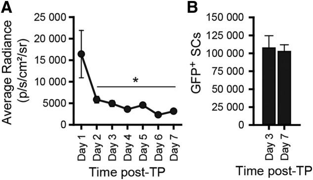
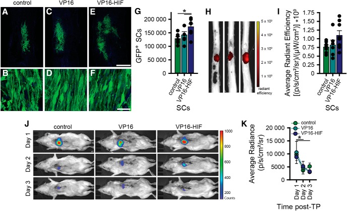
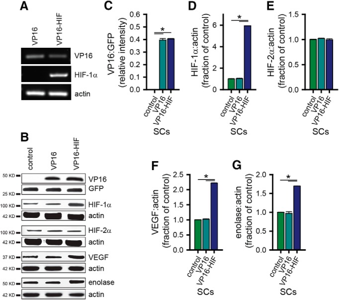
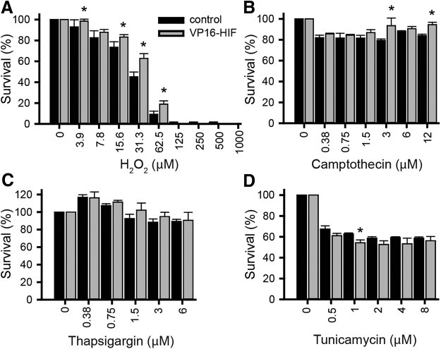
Cellular transplantation is in clinical testing for a number of central nervous system disorders, including spinal cord injury (SCI). One challenge is acute transplanted cell death. To prevent this death, there is a need to both establish when the death occurs and develop approaches to mitigate its effects. Here, using luciferase (luc) and green fluorescent protein (GFP) expressing Schwann cell (SC) transplants in the contused thoracic rat spinal cord 7 d postinjury, we establish via bioluminescent (IVIS) imaging and stereology that cell death occurs prior to 2-3 d postimplantation. We then test an alternative approach to the current paradigm of enhancing transplant survival by including multiple factors along with the cells. To stimulate multiple cellular adaptive pathways concurrently, we activate the hypoxia-inducible factor 1α (HIF-1α) transcriptional pathway. Retroviral expression of VP16-HIF-1α in SCs increased HIF-α by 5.9-fold and its target genes implicated in oxygen transport and delivery (VEGF, 2.2-fold) and cellular metabolism (enolase, 1.7-fold). In cell death assays , HIF-1α protected cells from HO-induced oxidative damage. It also provided some protection against camptothecin-induced DNA damage, but not thapsigargin-induced endoplasmic reticulum stress or tunicamycin-induced unfolded protein response. Following transplantation, VP16-HIF-1α increased SC survival by 34.3%. The increase in cell survival was detectable by stereology, but not by luciferase or GFP IVIS imaging. The results support the hypothesis that activating adaptive cellular pathways enhances transplant survival and identifies an alternative pro-survival approach that, with optimization, could be amenable to clinical translation.
细胞移植正在进行多项中枢神经系统疾病的临床测试,包括脊髓损伤(SCI)。一个挑战是急性移植细胞死亡。为了防止这种死亡,需要确定死亡发生的时间,并开发减轻其影响的方法。在这里,我们使用荧光素酶(luc)和绿色荧光蛋白(GFP)表达的施万细胞(SC)移植在损伤后 7 天的挫伤大鼠脊髓中,通过生物发光(IVIS)成像和立体学方法确定细胞死亡发生在植入后 2-3 天之前。然后,我们通过包括多种因子与细胞一起,测试了一种替代当前增强移植生存的范例的方法。为了同时激活多个细胞适应性途径,我们激活缺氧诱导因子 1α(HIF-1α)转录途径。SC 中的逆转录病毒表达 VP16-HIF-1α使 HIF-α增加 5.9 倍,其靶基因与氧运输和递送(VEGF,2.2 倍)和细胞代谢(烯醇酶,1.7 倍)有关。在细胞死亡测定中,HIF-1α保护细胞免受 HO 诱导的氧化损伤。它还为 camptothecin 诱导的 DNA 损伤提供了一些保护,但不能为 thapsigargin 诱导的内质网应激或 tunicamycin 诱导的未折叠蛋白反应提供保护。移植后,VP16-HIF-1α使 SC 存活率增加了 34.3%。通过立体学可以检测到细胞存活率的增加,但通过荧光素酶或 GFP IVIS 成像则无法检测到。这些结果支持这样的假设,即激活适应性细胞途径可增强移植的存活率,并确定了一种替代的生存促进方法,如果进行优化,可能适合临床转化。