Olsen Grethe M, Hovde Karoline, Kondo Hideki, Sakshaug Teri, Sømme Hanna Haaland, Whitlock Jonathan R, Witter Menno P
The Faculty of Medicine, Kavli Institute for Systems Neuroscience, Centre for Neural Computation, Egil and Pauline Braathen and Fred Kavli Centre for Cortical Microcircuits, NTNU-Norwegian University of Science and Technology, Trondheim, Norway.
Front Syst Neurosci. 2019 Aug 21;13:38. doi: 10.3389/fnsys.2019.00038. eCollection 2019.
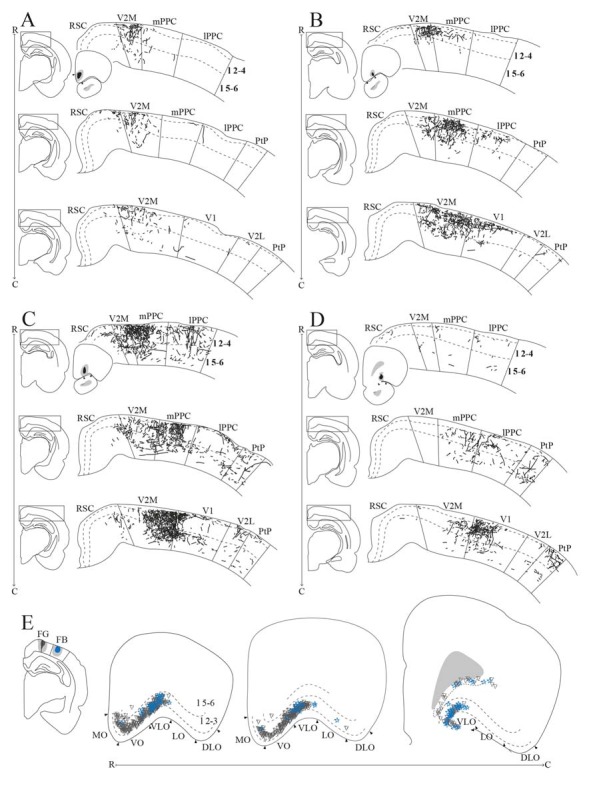
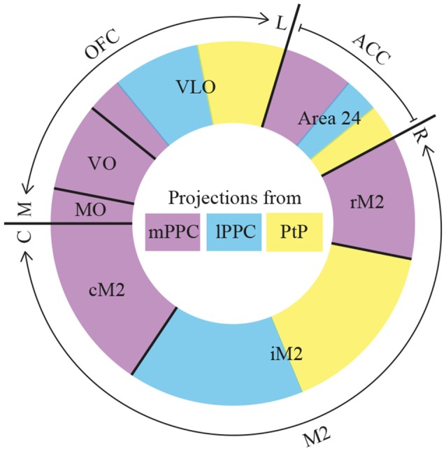
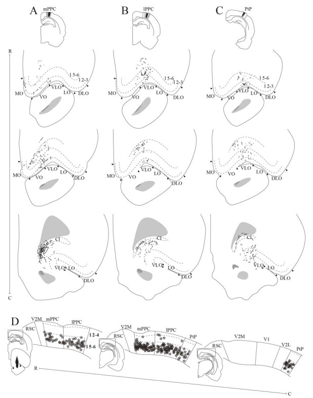
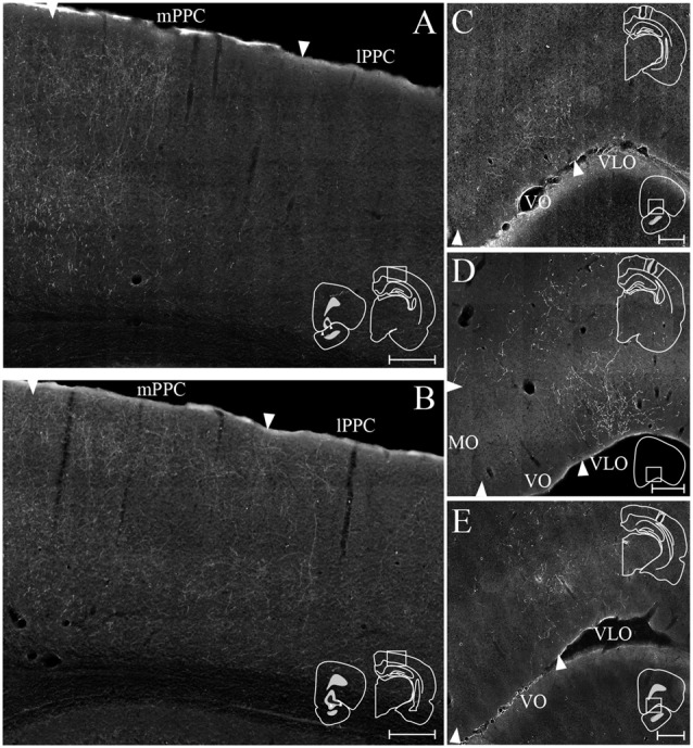
Recent investigations of the rat posterior parietal cortex (PPC) suggest that this region plays a central role in action control together with the frontal cortical areas. Posterior parietal-frontal cortical connections have been described in rats, but little is known about whether these connections are topographically organized as in the primate. Here, we injected retrograde and anterograde tracers into subdivisions of PPC as well as the frontal midline and orbital cortical areas to explore possible topographies within their connections. We found that PPC projects to several frontal cortical areas, largely reciprocating the densest input received from the same areas. All PPC subdivisions are strongly connected with the secondary motor cortex (M2) in a topographically organized manner. The medial subdivision (medial posterior parietal cortex, mPPC) has a dense reciprocal connection with the most caudal portion of M2 (cM2), whereas the lateral subdivision (lateral posterior parietal cortex, lPPC) and the caudolateral subdivision (PtP) are reciprocally connected with the intermediate rostrocaudal portion of M2 (iM2). Sparser reciprocal connections were seen with anterior cingulate area 24b. mPPC connects with rostral, and lPPC and PtP connect with caudal parts of 24b, respectively. There are virtually no connections with area 24a, nor with prelimbic or infralimbic cortex. PPC and orbitofrontal cortices are also connected, showing a gradient such that mPPC entertains reciprocal connections mainly with the ventral orbitofrontal cortex (OFC), whereas lPPC and PtP are preferentially connected with medial and central portions of ventrolateral OFC, respectively. Our results thus indicate that the connections of PPC with frontal cortices are organized in a topographical fashion, supporting functional heterogeneity within PPC and frontal cortices.
最近对大鼠后顶叶皮质(PPC)的研究表明,该区域与额叶皮质区域一起在动作控制中发挥核心作用。大鼠中已描述了后顶叶 - 额叶皮质连接,但对于这些连接是否像灵长类动物那样按拓扑结构组织却知之甚少。在此,我们将逆行和顺行示踪剂注入PPC的各个亚区以及额中线和眶额皮质区域,以探索它们连接中的可能拓扑结构。我们发现PPC投射到几个额叶皮质区域,在很大程度上与从相同区域接收的最密集输入相互对应。所有PPC亚区都以拓扑结构组织的方式与次级运动皮质(M2)紧密相连。内侧亚区(内侧后顶叶皮质,mPPC)与M2最尾端部分(cM2)有密集的相互连接,而外侧亚区(外侧后顶叶皮质,lPPC)和尾外侧亚区(PtP)与M2中间的头尾向部分(iM2)相互连接。与前扣带区24b的相互连接较为稀疏。mPPC与24b的头端相连,而lPPC和PtP分别与24b的尾端部分相连。实际上与24a区、前边缘皮质或边缘下皮质没有连接。PPC与眶额皮质也有连接,呈现出一种梯度,即mPPC主要与腹侧眶额皮质(OFC)有相互连接,而lPPC和PtP分别优先与腹外侧OFC的内侧和中央部分相连。因此,我们的结果表明PPC与额叶皮质的连接是以拓扑方式组织的,支持了PPC和额叶皮质内的功能异质性。