Roleder Tomasz, Kedhi Elvin, Berta Balazs, Gasior Pawel, Wanha Wojciech, Roleder Magda, Fluder Joanna, Smolka Grzegorz, Ochala Andrzej, Wojakowski Wojciech
Department of Cardiology and Structural Heart Diseases, 3 Division of Cardiology, Medical University of Silesia, Katowice, Poland.
Isala Haartcentrum, Zwolle, Netherlands.
Postepy Kardiol Interwencyjnej. 2019;15(2):143-150. doi: 10.5114/aic.2019.86009. Epub 2019 Jun 26.
To date the early strut coverage with the second-generation durable-polymer ONYX zotarolimus-eluting stent (O-ZES) is unknown.
Optical coherence tomography (OCT) assessed the strut coverage of O-ZES at thirty-day follow-up.
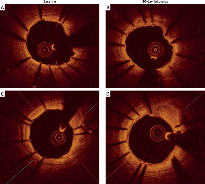
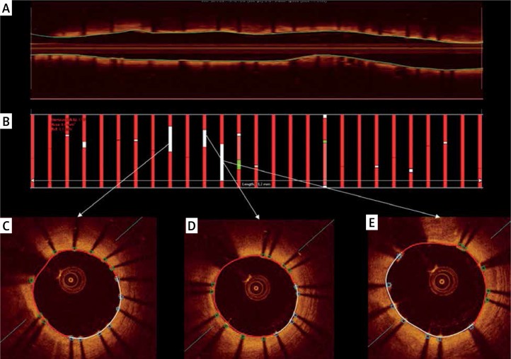
OCT was performed after implantation and at 1-month follow-up in 15 patients treated with O-ZES.
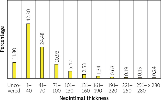
Mean patient age was 67 ±7 years (73% males). The clinical presentation consisted of acute coronary syndromes ( = 13) and stable coronary disease ( = 2). Four (26%) patients had diabetes. OCT analysis was performed at baseline and 1-month follow-up in all stents. 378 cross-sections with 3582 struts were assessed at baseline and 3661 at follow-up. At follow-up, 88% struts were covered by tissue with a median thickness 37.91 μm (IQR: 22.32-64.15). Median in-stent area obstruction by neointima was 2.64% (IQR: 1.70-4.84). From the total stent covered area, 92.3% showed complete strut coverage. Homogeneous tissue was observed in 74% of cases. There were no differences in minimal lumen area (5.07 ±1.08 mm vs. 4.81 ±0.94 mm, = 0.125) or minimal stent area (4.95 ±1.22 mm vs. 4.92 ±0.99 mm) at baseline and at follow-up. There were no differences in the rate of strut malapposition (4.3% vs. 5.7%, = 0.417). For all stents, malapposition volume was 47.9 mm at baseline and 51.7 mm at follow-up, giving the late acquired stent malapposition volume of 3.8 mm.
The second-generation durable polymer O-ZES showed favorable vessel healing at 30-day OCT follow-up.
迄今为止,第二代耐用聚合物佐他莫司洗脱支架(O-ZES)早期的支架小梁覆盖情况尚不清楚。
光学相干断层扫描(OCT)评估O-ZES在30天随访时的支架小梁覆盖情况。
对15例接受O-ZES治疗的患者在植入后及1个月随访时进行OCT检查。
患者平均年龄为67±7岁(73%为男性)。临床表现包括急性冠状动脉综合征(n = 13)和稳定型冠心病(n = 2)。4例(26%)患者患有糖尿病。对所有支架在基线和1个月随访时进行OCT分析。基线时评估了378个包含3582个支架小梁的横截面,随访时评估了3661个。随访时,88%的支架小梁被组织覆盖,组织厚度中位数为37.91μm(四分位间距:22.32 - 64.15)。新生内膜导致的支架内面积阻塞中位数为2.64%(四分位间距:1.70 - 4.84)。在整个支架覆盖区域中,92.3%显示支架小梁完全覆盖。74%的病例观察到组织均匀。基线和随访时最小管腔面积(5.07±1.08mm对4.81±0.94mm,P = 0.125)或最小支架面积(4.95±1.22mm对4.92±0.99mm)无差异。支架小梁贴壁不良率无差异(4.3%对5.7%,P = 0.417)。对于所有支架,基线时贴壁不良体积为47.9mm,随访时为51.7mm,晚期获得性支架贴壁不良体积为3.8mm。
第二代耐用聚合物O-ZES在OCT 30天随访时显示出良好的血管愈合情况。