Rolvien Tim, Krause Matthias, Zustin Jozef, Yastrebov Oleg, Oheim Ralf, Barvencik Florian, Frosch Karl-Heinz, Amling Michael
Department of Osteology and Biomechanics, University Medical Center Hamburg-Eppendorf, Hamburg, Germany.
Department of Orthopedics, University Medical Center Hamburg-Eppendorf, Hamburg, Germany.
J Bone Oncol. 2019 Aug 20;18:100256. doi: 10.1016/j.jbo.2019.100256. eCollection 2019 Oct.
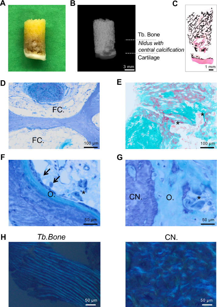
Osteoid osteoma (OO) is a benign bone tumor producing non-mineralized bone matrix (i.e., osteoid). While peritumoral edema is commonly found in OO, extensive bone marrow edema has been reported less frequently. Furthermore, the micro-morphological characteristics of the nidus and its central calcification remain unclear. In this study, a consecutive series of four patients suffering from extensive bone marrow edema triggered by intra-articular osteoid osteoma underwent clinical examination, magnetic resonance imaging (MRI) and computed tomography (CT) as well as dual-energy X-ray absorptiometry (DXA) and laboratory bone turnover analyses. The obtained resection specimens were processed by undecalcified histology and were subsequently analyzed by light microscopy and quantitative backscattered electron imaging (qBEI). We report an entity of intra-articular osteoid osteoma in the knee and foot, in which an extensive and persistent bone marrow edema syndrome masked the correct diagnosis. While metabolic bone diseases were excluded in all cases, the reassessment of the patients' clinical history including pain characteristics (nocturnal, aspirin sensitivity) led us to perform additional CT, where the tumor was diagnosed. The micro-morphological analysis of the OO biopsies revealed that the nidus was surrounded by hyperosteoidosis, while central mineralization was detected in all cases. This mineralized area showed a significantly higher mineralization heterogeneity than the surrounding trabecular bone and more disorganized collagen fibers detected by qBEI and polarized light microscopy, respectively. Taken together, our results indicate that osteoid osteoma should be considered when persistent and extensive, peri-articular bone marrow edema is diagnosed. The central calcification that is found inside the nidus in conventional imaging was mirrored by bone matrix with a heterogeneous mineralization pattern.
骨样骨瘤(OO)是一种产生非矿化骨基质(即类骨质)的良性骨肿瘤。虽然瘤周水肿在骨样骨瘤中很常见,但广泛的骨髓水肿报道较少。此外,瘤巢及其中心钙化的微观形态特征仍不清楚。在本研究中,对连续4例因关节内骨样骨瘤引发广泛骨髓水肿的患者进行了临床检查、磁共振成像(MRI)、计算机断层扫描(CT)以及双能X线吸收测定法(DXA)和实验室骨转换分析。所获得的切除标本采用不脱钙组织学方法处理,随后进行光学显微镜检查和定量背散射电子成像(qBEI)分析。我们报告了膝部和足部关节内骨样骨瘤的一种情况,其中广泛且持续的骨髓水肿综合征掩盖了正确诊断。虽然所有病例均排除了代谢性骨病,但对患者临床病史(包括疼痛特征,如夜间痛、阿司匹林敏感性)的重新评估使我们进行了额外的CT检查,从而诊断出肿瘤。对骨样骨瘤活检标本的微观形态分析显示,瘤巢被骨质增生包围,所有病例均检测到中心钙化。该矿化区域显示出比周围小梁骨明显更高的矿化异质性,并且分别通过qBEI和偏振光显微镜检测到更紊乱的胶原纤维。综上所述,我们的结果表明,当诊断出持续且广泛的关节周围骨髓水肿时,应考虑骨样骨瘤。传统成像中在瘤巢内发现的中心钙化在具有异质矿化模式的骨基质中得到反映。