Baccouche Khadija, Fakhfekh Rym, Khalifa Dhouha, Hachfi Haifa, Daldoul Cyrine, Elamri Nejla, Bouajina Elyes
Rheumatology Department of Farhat Hached University Hospital, Faculty of Medicine of Sousse, University of Sousse, Sousse, Tunisia.
Case Rep Med. 2025 Apr 28;2025:7648066. doi: 10.1155/carm/7648066. eCollection 2025.
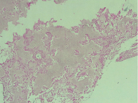
Osteoid osteomas predominantly occur in the cortices of long bones, with the femur and tibia being the most commonly affected sites. However, they can occasionally present in atypical locations, such as the carpus, which can lead to diagnostic confusion with other conditions. This case report details an intraarticular osteoid osteoma in the trapezoid bone. Initial evaluations, including standard radiographs, joint ultrasound, and wrist MRI performed twice, initially pointed toward a diagnosis of wrist synovitis. This case underscores the diagnostic challenges posed by atypical presentations of osteoid osteomas. Given the edema present in the carpal bones alongside the synovitis, we performed a hand CT scan, which raised doubts about the appearance of the nidus and histopathological examination confirmed the diagnosis. Clinical symptoms, including pain and functional limitations, were completely resolved following surgical excision.
骨样骨瘤主要发生于长骨皮质,其中股骨和胫骨是最常受累部位。然而,它们偶尔也会出现在非典型部位,如腕骨,这可能导致与其他疾病的诊断混淆。本病例报告详细介绍了一例梯形骨内的关节内骨样骨瘤。最初的评估,包括标准X线片、关节超声以及两次腕关节MRI检查,最初指向腕滑膜炎的诊断。本病例强调了骨样骨瘤非典型表现所带来的诊断挑战。鉴于腕骨出现水肿并伴有滑膜炎,我们进行了手部CT扫描,这对瘤巢的表现提出了疑问,而组织病理学检查确诊了该病。手术切除后,包括疼痛和功能受限在内的临床症状完全缓解。