• 文献检索
  • 文档翻译
  • 深度研究
  • 学术资讯
  • Suppr Zotero 插件Zotero 插件
  • 邀请有礼
  • 套餐&价格
  • 历史记录
应用&插件
Suppr Zotero 插件Zotero 插件浏览器插件Mac 客户端Windows 客户端微信小程序
定价
高级版会员购买积分包购买API积分包
服务
文献检索文档翻译深度研究API 文档MCP 服务
关于我们
关于 Suppr公司介绍联系我们用户协议隐私条款
关注我们

Suppr 超能文献

核心技术专利:CN118964589B侵权必究
粤ICP备2023148730 号-1Suppr @ 2026

文献检索

告别复杂PubMed语法,用中文像聊天一样搜索,搜遍4000万医学文献。AI智能推荐,让科研检索更轻松。

立即免费搜索

文件翻译

保留排版,准确专业,支持PDF/Word/PPT等文件格式,支持 12+语言互译。

免费翻译文档

深度研究

AI帮你快速写综述,25分钟生成高质量综述,智能提取关键信息,辅助科研写作。

立即免费体验

高b值扩散加权成像在前列腺癌检测中的信号强度

Signal Intensity of High B-value Diffusion-weighted Imaging for the Detection of Prostate Cancer.

作者信息

A Rezaeian, M J Tahmasebi Birgani, N Chegeni, M Sarkarian, M Gh Hanafi, Gh Akbarizadeh

机构信息

Department of Medical Physics, Faculty of Medicine, Ahvaz Jundishapur University of Medical Sciences, Ahvaz, Iran.

Department of Urology, Golestan Hospital, Ahvaz Jundishapur University of Medical Sciences, Ahvaz, Iran.

出版信息

J Biomed Phys Eng. 2019 Aug 1;9(4):453-458. doi: 10.31661/jbpe.v0i0.811. eCollection 2019 Aug.

DOI:10.31661/jbpe.v0i0.811
PMID:31531298
原文链接:https://pmc.ncbi.nlm.nih.gov/articles/PMC6709361/
Abstract

BACKGROUND

Diffusion-weighted imaging (DWI) is a main component of multiparametric MRI for prostate cancer detection. Recently, high b value DWI has gained more attention because of its capability for tumor characterization.

OBJECTIVE

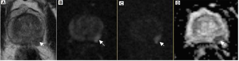
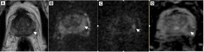
To assess based on histopathological findings of transrectal ultrasound (TRUS)-guided prostate biopsy as a reference, an increase in signal intensity of prostatic lesions in comparison with normal background tissue on high b-value diffusion-weighted images could be a sign of malignancy.

MATERIAL AND METHODS

Fifty-three consecutive patients retrospectively included in the study. All patients underwent routine TRUS-guided prostate biopsies involving 12 cores after the magnetic resonance imaging (MRI) examinations. In seventeen patients (n =35 lesions), the prostate cancer was histologically confirmed by TRUS-guided prostate biopsy. The biopsy results of other patients were negative. Signal intensities on the high b-value (1600 s/mm) images of the peripheral zone, the central gland, and the defined lesions were evaluated using region of interest-based measurements. Sensitivity, specificity, positive predictive value (PPV) and negative predictive value (NPV) for prostate cancer detection using signal intensity of high b value diffusion-weighted images were calculated.

RESULTS

In the patients with confirmed prostate cancer, fourteen had visually increased SI on the high b-value images. The SI of lesions for these patients was higher than the SI of peripheral zone (22±18%) or central gland (31±20%). In patients with a negative biopsy, eight had visually increased SI on the high b-value images. The SI of lesions for these patients was 23±21% and 35±18% higher than the SI in the peripheral zone and the central gland, respectively. The sensitivity, specificity, PPV, and NPV for prostate cancer using SI of high b value DWI were 71, 87, 62, and 87 %, respectively.

CONCLUSION

Visually increased SI on the high b-value images can be an indication of malignancy, although some benign lesions also show this increase in signal intensity.

摘要

背景

扩散加权成像(DWI)是多参数MRI用于前列腺癌检测的主要组成部分。近年来,高b值DWI因其具有肿瘤特征化能力而受到更多关注。

目的

以经直肠超声(TRUS)引导下前列腺穿刺活检的组织病理学结果为参考,评估高b值扩散加权图像上前列腺病变与正常背景组织相比信号强度增加是否可能提示恶性肿瘤。

材料与方法

回顾性纳入连续53例患者。所有患者在磁共振成像(MRI)检查后均接受了常规TRUS引导下的前列腺穿刺活检,共取12针组织。17例患者(n = 35个病变)经TRUS引导下前列腺穿刺活检组织学确诊为前列腺癌。其他患者的活检结果为阴性。使用基于感兴趣区的测量方法评估外周带、中央腺体及明确病变在高b值(1600 s/mm²)图像上的信号强度。计算使用高b值扩散加权图像信号强度检测前列腺癌的敏感性、特异性、阳性预测值(PPV)和阴性预测值(NPV)。

结果

在确诊为前列腺癌的患者中,14例在高b值图像上可见信号强度增加。这些患者病变的信号强度高于外周带(22±18%)或中央腺体(31±20%)。活检结果为阴性的患者中,8例在高b值图像上可见信号强度增加。这些患者病变的信号强度分别比外周带和中央腺体的信号强度高23±21%和35±18%。使用高b值DWI信号强度检测前列腺癌的敏感性、特异性、PPV和NPV分别为71%、87%、62%和87%。

结论

高b值图像上信号强度明显增加可能提示恶性肿瘤,尽管一些良性病变也会出现这种信号强度增加。

https://cdn.ncbi.nlm.nih.gov/pmc/blobs/c676/6709361/43d90bba3121/JBPE-9-453-g002.jpg
https://cdn.ncbi.nlm.nih.gov/pmc/blobs/c676/6709361/c352e00b1098/JBPE-9-453-g001.jpg
https://cdn.ncbi.nlm.nih.gov/pmc/blobs/c676/6709361/43d90bba3121/JBPE-9-453-g002.jpg
https://cdn.ncbi.nlm.nih.gov/pmc/blobs/c676/6709361/c352e00b1098/JBPE-9-453-g001.jpg
https://cdn.ncbi.nlm.nih.gov/pmc/blobs/c676/6709361/43d90bba3121/JBPE-9-453-g002.jpg

相似文献

1
Signal Intensity of High B-value Diffusion-weighted Imaging for the Detection of Prostate Cancer.高b值扩散加权成像在前列腺癌检测中的信号强度
J Biomed Phys Eng. 2019 Aug 1;9(4):453-458. doi: 10.31661/jbpe.v0i0.811. eCollection 2019 Aug.
2
Increased signal intensity of prostate lesions on high b-value diffusion-weighted images as a predictive sign of malignancy.高 b 值扩散加权图像上前列腺病变信号强度增高是恶性病变的预测征象。
Eur Radiol. 2014 Jan;24(1):209-13. doi: 10.1007/s00330-013-2999-3. Epub 2013 Aug 31.
3
Accuracy of Diffusion Weighted Images and MR Spectroscopy in Prostate Lesions - Our Experience with Endorectal Coil on 1.5 T MRI.扩散加权成像和磁共振波谱在前列腺病变中的准确性——我们在1.5T磁共振成像上使用直肠内线圈的经验
J Clin Diagn Res. 2017 May;11(5):TC10-TC14. doi: 10.7860/JCDR/2017/27754.9825. Epub 2017 May 1.
4
Multiparametric MRI in detection and staging of prostate cancer.多参数磁共振成像在前列腺癌检测与分期中的应用
Dan Med J. 2017 Feb;64(2).
5
Diffusion-weighted MRI for locally recurrent prostate cancer after external beam radiotherapy.扩散加权 MRI 在外照射放疗后局部复发性前列腺癌的应用。
AJR Am J Roentgenol. 2012 Mar;198(3):596-602. doi: 10.2214/AJR.11.7162.
6
Evaluation of Diffusion Kurtosis Imaging Versus Standard Diffusion Imaging for Detection and Grading of Peripheral Zone Prostate Cancer.扩散峰度成像与标准扩散成像在周围区前列腺癌检测及分级中的评估
Invest Radiol. 2015 Aug;50(8):483-9. doi: 10.1097/RLI.0000000000000155.
7
Multiparametric MRI to improve detection of prostate cancer compared with transrectal ultrasound-guided prostate biopsy alone: the PROMIS study.多参数 MRI 提高经直肠超声引导前列腺活检单独诊断前列腺癌的检出率:PROMIS 研究。
Health Technol Assess. 2018 Jul;22(39):1-176. doi: 10.3310/hta22390.
8
[Effects of different computed high b-values on diffusion weighted imaging scores in Prostate Imaging Reporting and Data System version 2 of prostate cancer in peripheral zone].[不同计算高b值对前列腺影像报告和数据系统第2版外周带前列腺癌扩散加权成像评分的影响]
Zhonghua Yi Xue Za Zhi. 2017 Nov 21;97(43):3401-3405. doi: 10.3760/cma.j.issn.0376-2491.2017.43.009.
9
Predicting Prostate Biopsy Outcomes: A Preliminary Investigation on Screening with Ultrahigh B-Value Diffusion-Weighted Imaging as an Innovative Diagnostic Biomarker.预测前列腺活检结果:超高 b 值扩散加权成像作为一种创新诊断生物标志物进行筛查的初步研究。
PLoS One. 2016 Mar 10;11(3):e0151176. doi: 10.1371/journal.pone.0151176. eCollection 2016.
10
Negative predictive value of multiparametric MRI for prostate cancer detection: outcome of 5-year follow-up in men with negative findings on initial MRI studies.多参数磁共振成像对前列腺癌检测的阴性预测值:初始磁共振成像研究结果为阴性的男性患者5年随访结果
Eur J Radiol. 2014 Oct;83(10):1740-5. doi: 10.1016/j.ejrad.2014.06.026. Epub 2014 Jul 3.

引用本文的文献

1
Accelerated diffusion-weighted imaging of the prostate employing echo planar imaging with compressed SENSE based reconstruction.采用基于压缩感知重建的回波平面成像的前列腺加速扩散加权成像。
Sci Rep. 2025 Mar 25;15(1):10265. doi: 10.1038/s41598-025-94777-6.
2
Diffusion-weighted magnetic resonance imaging at 1.5 T for peripheral zone prostate cancer: the influence of the -value combination on the diagnostic performance of apparent diffusion coefficient.1.5T磁共振扩散加权成像在外周带前列腺癌中的应用:b值组合对表观扩散系数诊断效能的影响
Pol J Radiol. 2022 Apr 8;87:e215-e219. doi: 10.5114/pjr.2022.115715. eCollection 2022.
3

本文引用的文献

1
Optimal high b-value for diffusion weighted MRI in diagnosing high risk prostate cancers in the peripheral zone.扩散加权磁共振成像诊断外周带高危前列腺癌的最佳高b值
J Magn Reson Imaging. 2017 Jan;45(1):125-131. doi: 10.1002/jmri.25353. Epub 2016 Jul 7.
2
Predicting Prostate Biopsy Outcomes: A Preliminary Investigation on Screening with Ultrahigh B-Value Diffusion-Weighted Imaging as an Innovative Diagnostic Biomarker.预测前列腺活检结果:超高 b 值扩散加权成像作为一种创新诊断生物标志物进行筛查的初步研究。
PLoS One. 2016 Mar 10;11(3):e0151176. doi: 10.1371/journal.pone.0151176. eCollection 2016.
3
Diffusion in prostate cancer detection on a 3T scanner: How many b-values are needed?
Detection of High-Grade Prostate Cancer With a Super High B-value (4000 s/mm2) in Diffusion-Weighted Imaging Sequences by Magnetic Resonance Imaging.
磁共振成像扩散加权成像序列中超高b值(4000 s/mm2)检测高级别前列腺癌
Cureus. 2022 Mar 3;14(3):e22807. doi: 10.7759/cureus.22807. eCollection 2022 Mar.
3T磁共振成像仪在前列腺癌检测中的扩散加权成像:需要多少个b值?
J Magn Reson Imaging. 2016 Sep;44(3):601-9. doi: 10.1002/jmri.25206. Epub 2016 Feb 23.
4
Apparent diffusion coefficient value is a strong predictor of unsuspected aggressiveness of prostate cancer before radical prostatectomy.表观扩散系数值是前列腺癌根治术前未被怀疑的侵袭性的有力预测指标。
World J Urol. 2016 Oct;34(10):1389-95. doi: 10.1007/s00345-016-1789-3. Epub 2016 Feb 22.
5
PI-RADS Prostate Imaging - Reporting and Data System: 2015, Version 2.PI-RADS前列腺影像报告和数据系统:2015版,第2版
Eur Urol. 2016 Jan;69(1):16-40. doi: 10.1016/j.eururo.2015.08.052. Epub 2015 Oct 1.
6
Changes in Epithelium, Stroma, and Lumen Space Correlate More Strongly with Gleason Pattern and Are Stronger Predictors of Prostate ADC Changes than Cellularity Metrics.上皮、基质和管腔空间的变化与 Gleason 模式的相关性更强,并且比细胞计数指标更能有力地预测前列腺 ADC 的变化。
Radiology. 2015 Dec;277(3):751-62. doi: 10.1148/radiol.2015142414. Epub 2015 Jun 23.
7
Diffusion-weighted magnetic resonance imaging for prediction of insignificant prostate cancer in potential candidates for active surveillance.扩散加权磁共振成像用于预测主动监测潜在候选者中低危前列腺癌
Eur Radiol. 2015 Jun;25(6):1786-92. doi: 10.1007/s00330-014-3566-2. Epub 2015 Jan 31.
8
Comparison of calculated and acquired high b value diffusion-weighted imaging in prostate cancer.前列腺癌中计算得出的与采集到的高b值扩散加权成像的比较。
Abdom Imaging. 2015 Mar;40(3):578-86. doi: 10.1007/s00261-014-0246-2.
9
High and ultra-high b-value diffusion-weighted imaging in prostate cancer: a quantitative analysis.前列腺癌的高b值和超高b值扩散加权成像:定量分析
Acta Radiol. 2015 Aug;56(8):1009-15. doi: 10.1177/0284185114547900. Epub 2014 Aug 28.
10
Quantitative evaluation of computed high B value diffusion-weighted magnetic resonance imaging of the prostate.前列腺计算机高B值扩散加权磁共振成像的定量评估
Invest Radiol. 2013 Nov;48(11):779-86. doi: 10.1097/RLI.0b013e31829705bb.