Center for Medical Physics and Biomedical Engineering, Medical University of Vienna, Währinger Gürtel 18-20, A-1090 Vienna, Austria.
Christian Doppler Laboratory for Ocular and Dermal Effects of Thiomers, Medical University of Vienna, Währinger Gürtel 18-20, A-1090 Vienna, Austria.
Sci Rep. 2019 Sep 20;9(1):13643. doi: 10.1038/s41598-019-50104-4.
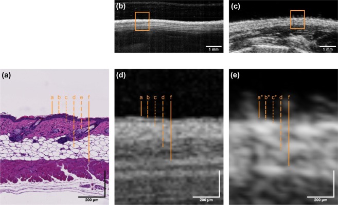
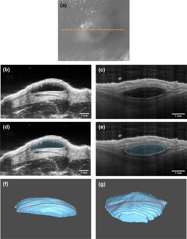
Optical coherence tomography (OCT) and high-frequency ultrasound (HFUS), two established imaging modalities in the field of dermatology, were evaluated and compared regarding their applicability for visualization of skin tissue morphology and quantification of murine intradermal structures. The accuracy and reproducibility of both methods were assessed ex vivo and in vivo using a standardized model for intradermal volumes based on injected soft tissue fillers. OCT revealed greater detail in skin morphology, allowing for detection of single layers due to the superior resolution. Volumetric data measured by OCT (7.9 ± 0.3 μl) and HFUS (7.7 ± 0.5 μl) were in good agreement and revealed a high accuracy when compared to the injected volume of 7.98 ± 0.8 µl. In vivo, OCT provided a higher precision (relative SD: 26% OCT vs. 42% HFUS) for the quantification of intradermal structures, whereas HFUS offered increased penetration depth enabling the visualization of deeper structures. A combination of both imaging technologies might be valuable for tumor assessments or other dermal pathologies in clinical settings.
光学相干断层扫描 (OCT) 和高频超声 (HFUS) 是皮肤科领域中两种成熟的成像方式,本研究旨在评估和比较这两种方法在可视化皮肤组织形态和量化鼠类皮内结构方面的适用性。通过基于注射软组织填充剂的标准化皮内容积模型,在体外用和离体评估了这两种方法的准确性和可重复性。OCT 可更详细地显示皮肤形态,由于其具有更高的分辨率,甚至可以检测到单层结构。OCT 测量的容积数据(7.9 ± 0.3 μl)和 HFUS(7.7 ± 0.5 μl)与注射体积 7.98 ± 0.8 μl 非常吻合,具有很高的准确性。在体内,OCT 对皮内结构的定量具有更高的精度(相对标准偏差:26% OCT 比 42% HFUS),而 HFUS 则具有更高的穿透深度,能够可视化更深的结构。这两种成像技术的结合可能对临床环境中的肿瘤评估或其他皮肤病变具有重要价值。